Iron »
PDB 6xaj-6xz8 »
6xig »
Iron in PDB 6xig: X-Ray Crystal Structure of Mqne From Pedobacter Heparinus
Enzymatic activity of X-Ray Crystal Structure of Mqne From Pedobacter Heparinus
All present enzymatic activity of X-Ray Crystal Structure of Mqne From Pedobacter Heparinus:
2.5.1.120;
2.5.1.120;
Protein crystallography data
The structure of X-Ray Crystal Structure of Mqne From Pedobacter Heparinus, PDB code: 6xig
was solved by
T.L.Grove,
J.B.Bonanno,
S.C.Almo,
with X-Ray Crystallography technique. A brief refinement statistics is given in the table below:
| Resolution Low / High (Å) | 19.92 / 1.59 |
| Space group | P 1 21 1 |
| Cell size a, b, c (Å), α, β, γ (°) | 72.968, 75.403, 83.921, 90.00, 107.69, 90.00 |
| R / Rfree (%) | 15.6 / 18.3 |
Iron Binding Sites:
The binding sites of Iron atom in the X-Ray Crystal Structure of Mqne From Pedobacter Heparinus
(pdb code 6xig). This binding sites where shown within
5.0 Angstroms radius around Iron atom.
In total 8 binding sites of Iron where determined in the X-Ray Crystal Structure of Mqne From Pedobacter Heparinus, PDB code: 6xig:
Jump to Iron binding site number: 1; 2; 3; 4; 5; 6; 7; 8;
In total 8 binding sites of Iron where determined in the X-Ray Crystal Structure of Mqne From Pedobacter Heparinus, PDB code: 6xig:
Jump to Iron binding site number: 1; 2; 3; 4; 5; 6; 7; 8;
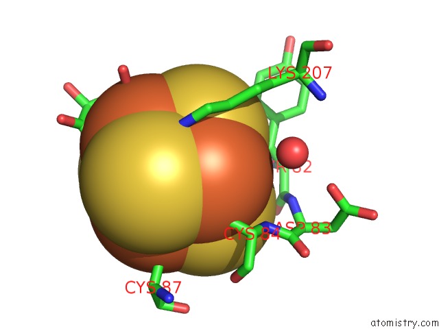
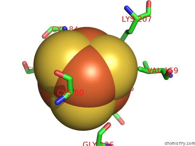
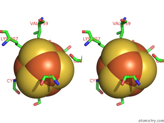
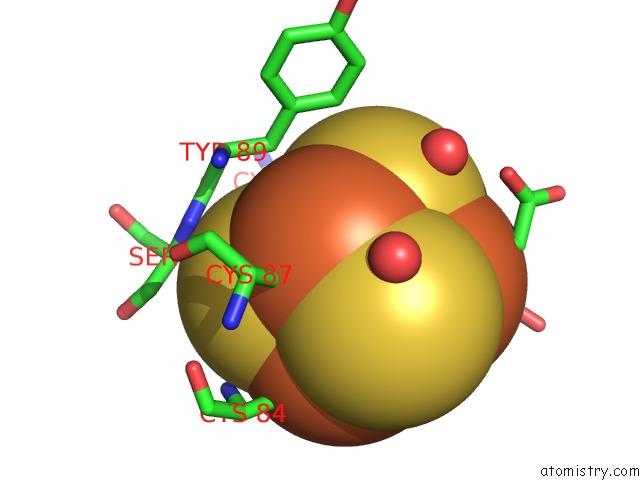
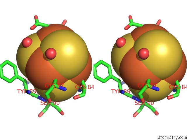
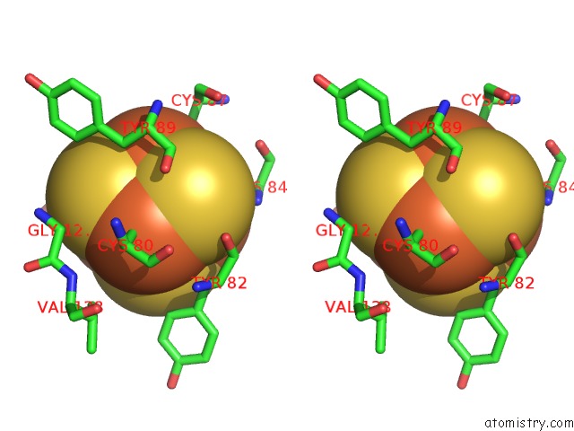
Iron binding site 1 out of 8 in 6xig
Go back to
Iron binding site 1 out
of 8 in the X-Ray Crystal Structure of Mqne From Pedobacter Heparinus
Mono view
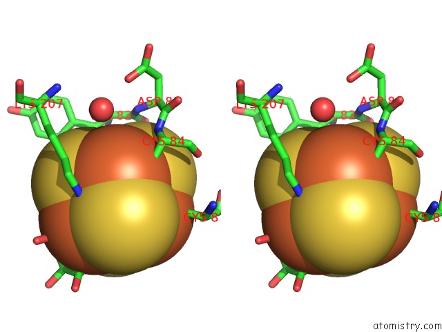
Stereo pair view
Mono view
Stereo pair view
A full contact list of Iron with other atoms in the Fe binding
site number 1 of X-Ray Crystal Structure of Mqne From Pedobacter Heparinus within 5.0Å range:
|
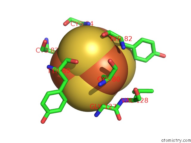
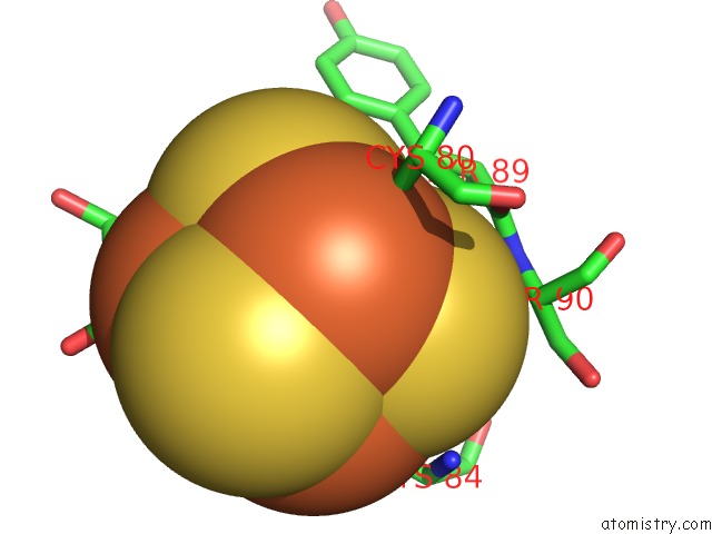
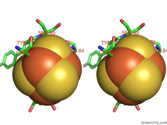
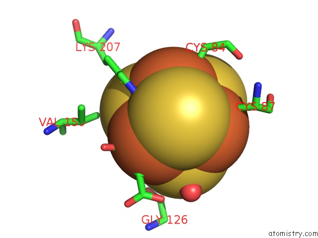
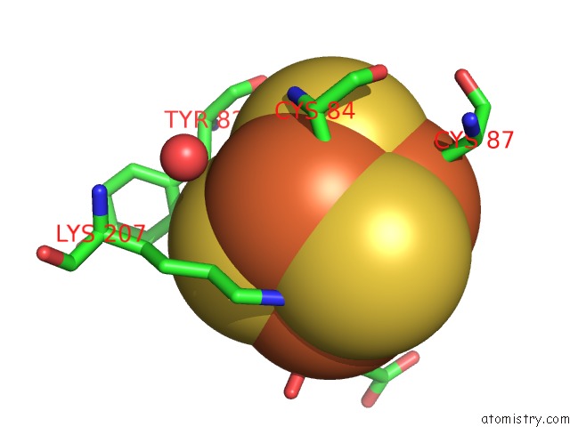
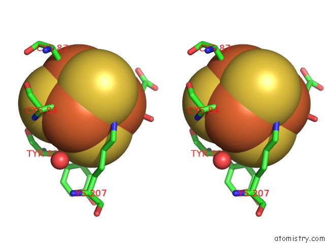
Iron binding site 2 out of 8 in 6xig
Go back to
Iron binding site 2 out
of 8 in the X-Ray Crystal Structure of Mqne From Pedobacter Heparinus
Mono view
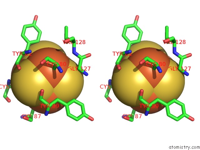
Stereo pair view
Mono view
Stereo pair view
A full contact list of Iron with other atoms in the Fe binding
site number 2 of X-Ray Crystal Structure of Mqne From Pedobacter Heparinus within 5.0Å range:
|
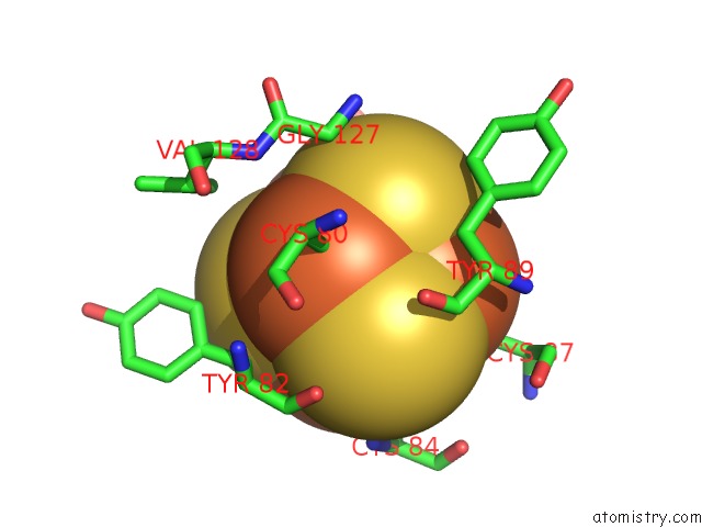
Iron binding site 3 out of 8 in 6xig
Go back to
Iron binding site 3 out
of 8 in the X-Ray Crystal Structure of Mqne From Pedobacter Heparinus
Mono view
Stereo pair view
Mono view
Stereo pair view
A full contact list of Iron with other atoms in the Fe binding
site number 3 of X-Ray Crystal Structure of Mqne From Pedobacter Heparinus within 5.0Å range:
|
Iron binding site 4 out of 8 in 6xig
Go back to
Iron binding site 4 out
of 8 in the X-Ray Crystal Structure of Mqne From Pedobacter Heparinus
Mono view
Stereo pair view
Mono view
Stereo pair view
A full contact list of Iron with other atoms in the Fe binding
site number 4 of X-Ray Crystal Structure of Mqne From Pedobacter Heparinus within 5.0Å range:
|
Iron binding site 5 out of 8 in 6xig
Go back to
Iron binding site 5 out
of 8 in the X-Ray Crystal Structure of Mqne From Pedobacter Heparinus
Mono view
Stereo pair view
Mono view
Stereo pair view
A full contact list of Iron with other atoms in the Fe binding
site number 5 of X-Ray Crystal Structure of Mqne From Pedobacter Heparinus within 5.0Å range:
|
Iron binding site 6 out of 8 in 6xig
Go back to
Iron binding site 6 out
of 8 in the X-Ray Crystal Structure of Mqne From Pedobacter Heparinus
Mono view

Stereo pair view
Mono view
Stereo pair view
A full contact list of Iron with other atoms in the Fe binding
site number 6 of X-Ray Crystal Structure of Mqne From Pedobacter Heparinus within 5.0Å range:
|
Iron binding site 7 out of 8 in 6xig
Go back to
Iron binding site 7 out
of 8 in the X-Ray Crystal Structure of Mqne From Pedobacter Heparinus
Mono view
Stereo pair view
Mono view
Stereo pair view
A full contact list of Iron with other atoms in the Fe binding
site number 7 of X-Ray Crystal Structure of Mqne From Pedobacter Heparinus within 5.0Å range:
|
Iron binding site 8 out of 8 in 6xig
Go back to
Iron binding site 8 out
of 8 in the X-Ray Crystal Structure of Mqne From Pedobacter Heparinus
Mono view
Stereo pair view
Mono view
Stereo pair view
A full contact list of Iron with other atoms in the Fe binding
site number 8 of X-Ray Crystal Structure of Mqne From Pedobacter Heparinus within 5.0Å range:
|
Reference:
A.G.Carl,
L.D.Harris,
M.Feng,
L.U.Nordstrom,
G.J.Gerfen,
G.B.Evans,
A.Silakov,
S.C.Almo,
T.L.Grove.
Narrow-Spectrum Antibiotic Targeting of the Radical Sam Enzyme Mqne in Menaquinone Biosynthesis. Biochemistry V. 59 2562 2020.
ISSN: ISSN 0006-2960
PubMed: 32627538
DOI: 10.1021/ACS.BIOCHEM.0C00070
Page generated: Wed Aug 6 15:55:34 2025
ISSN: ISSN 0006-2960
PubMed: 32627538
DOI: 10.1021/ACS.BIOCHEM.0C00070
Last articles
Fe in 6ZKLFe in 6ZKI
Fe in 6ZKK
Fe in 6ZKJ
Fe in 6ZKH
Fe in 6ZKG
Fe in 6ZKF
Fe in 6ZKE
Fe in 6ZKD
Fe in 6ZKC