Iron »
PDB 6xaj-6xz8 »
6xtf »
Iron in PDB 6xtf: Crystal Structure A Thioredoxin Reductase From Gloeobacter Violaceus Bound to Its Electron Donor
Protein crystallography data
The structure of Crystal Structure A Thioredoxin Reductase From Gloeobacter Violaceus Bound to Its Electron Donor, PDB code: 6xtf
was solved by
R.M.Buey,
G.Gonzalez-Holgado,
D.Fernandez-Justel,
M.Balsera,
with X-Ray Crystallography technique. A brief refinement statistics is given in the table below:
| Resolution Low / High (Å) | 83.37 / 2.23 |
| Space group | C 2 2 21 |
| Cell size a, b, c (Å), α, β, γ (°) | 166.741, 181.312, 80.369, 90, 90, 90 |
| R / Rfree (%) | 20.1 / 23.2 |
Iron Binding Sites:
The binding sites of Iron atom in the Crystal Structure A Thioredoxin Reductase From Gloeobacter Violaceus Bound to Its Electron Donor
(pdb code 6xtf). This binding sites where shown within
5.0 Angstroms radius around Iron atom.
In total 4 binding sites of Iron where determined in the Crystal Structure A Thioredoxin Reductase From Gloeobacter Violaceus Bound to Its Electron Donor, PDB code: 6xtf:
Jump to Iron binding site number: 1; 2; 3; 4;
In total 4 binding sites of Iron where determined in the Crystal Structure A Thioredoxin Reductase From Gloeobacter Violaceus Bound to Its Electron Donor, PDB code: 6xtf:
Jump to Iron binding site number: 1; 2; 3; 4;
Iron binding site 1 out of 4 in 6xtf
Go back to
Iron binding site 1 out
of 4 in the Crystal Structure A Thioredoxin Reductase From Gloeobacter Violaceus Bound to Its Electron Donor

Mono view
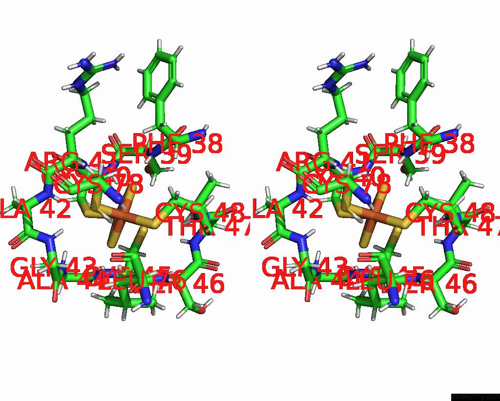
Stereo pair view
Mono view
Stereo pair view
A full contact list of Iron with other atoms in the Fe binding
site number 1 of Crystal Structure A Thioredoxin Reductase From Gloeobacter Violaceus Bound to Its Electron Donor within 5.0Å range:
|
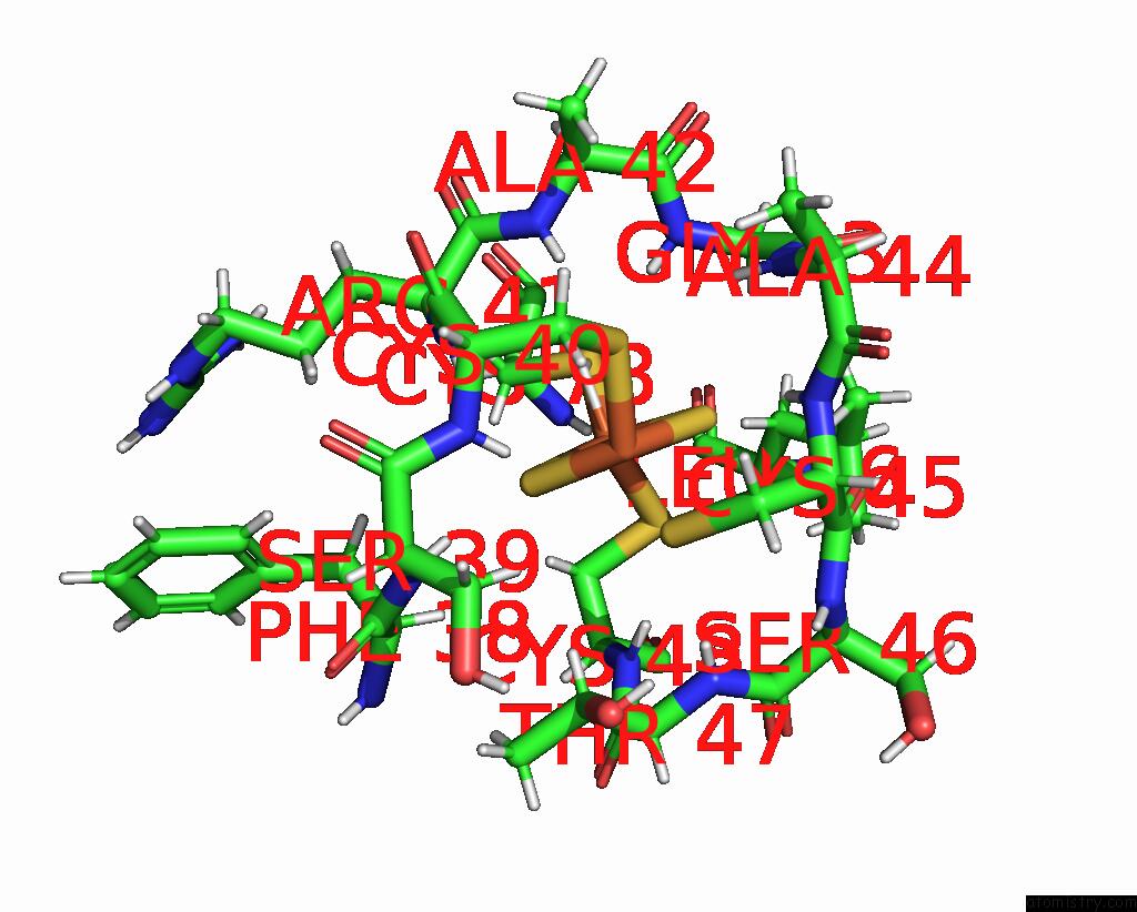
Iron binding site 2 out of 4 in 6xtf
Go back to
Iron binding site 2 out
of 4 in the Crystal Structure A Thioredoxin Reductase From Gloeobacter Violaceus Bound to Its Electron Donor

Mono view

Stereo pair view
Mono view
Stereo pair view
A full contact list of Iron with other atoms in the Fe binding
site number 2 of Crystal Structure A Thioredoxin Reductase From Gloeobacter Violaceus Bound to Its Electron Donor within 5.0Å range:
|
Iron binding site 3 out of 4 in 6xtf
Go back to
Iron binding site 3 out
of 4 in the Crystal Structure A Thioredoxin Reductase From Gloeobacter Violaceus Bound to Its Electron Donor
Mono view
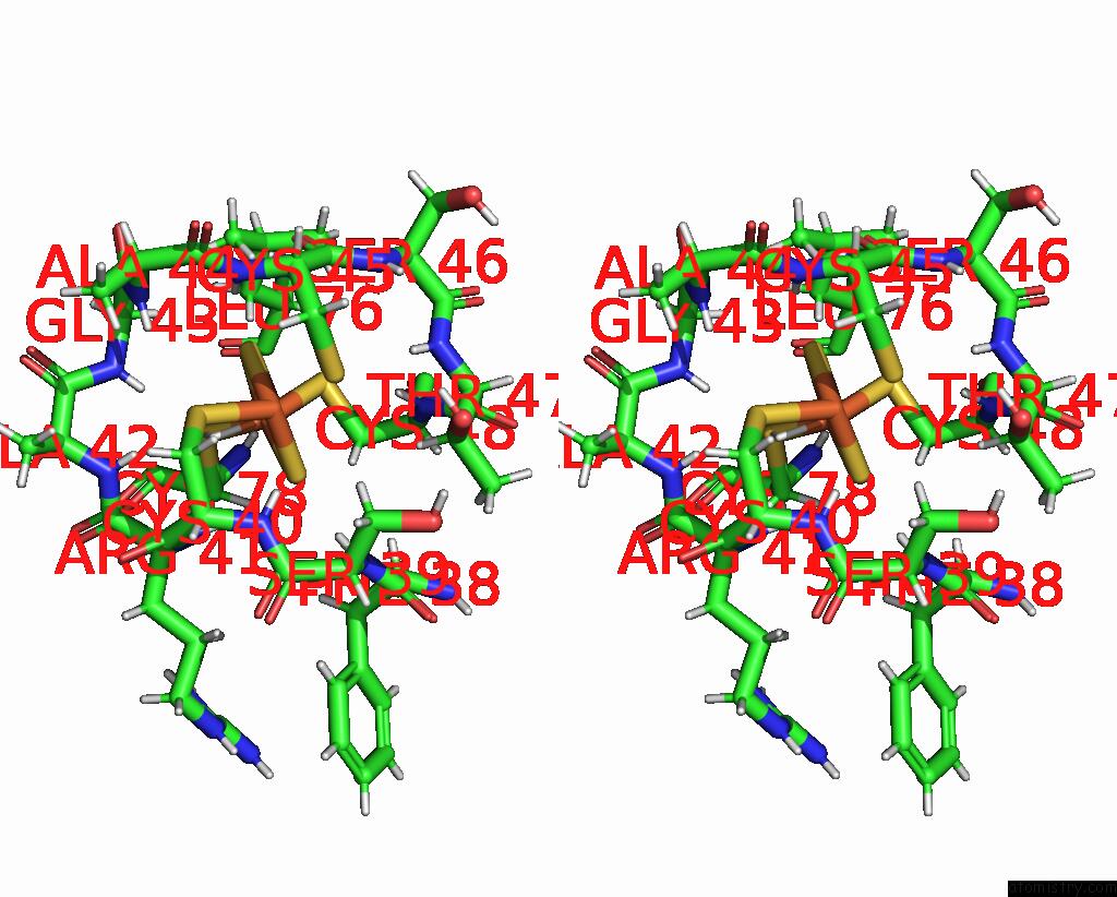
Stereo pair view
Mono view
Stereo pair view
A full contact list of Iron with other atoms in the Fe binding
site number 3 of Crystal Structure A Thioredoxin Reductase From Gloeobacter Violaceus Bound to Its Electron Donor within 5.0Å range:
|
Iron binding site 4 out of 4 in 6xtf
Go back to
Iron binding site 4 out
of 4 in the Crystal Structure A Thioredoxin Reductase From Gloeobacter Violaceus Bound to Its Electron Donor
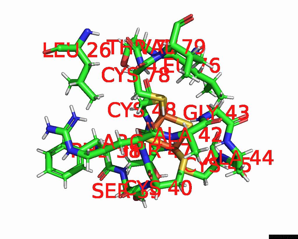
Mono view

Stereo pair view
Mono view
Stereo pair view
A full contact list of Iron with other atoms in the Fe binding
site number 4 of Crystal Structure A Thioredoxin Reductase From Gloeobacter Violaceus Bound to Its Electron Donor within 5.0Å range:
|
Reference:
R.M.Buey,
G.Gonzalez-Holgado,
D.Fernandez-Justel,
M.Balsera.
Crystal Structure A Thioredoxin Reductase From Gloeobacter Violaceus Bound to Its Electron Donor To Be Published.
Page generated: Wed Aug 6 16:03:50 2025
Last articles
Fe in 6ZVPFe in 6ZTW
Fe in 6ZTX
Fe in 6ZOO
Fe in 6ZTV
Fe in 6ZSK
Fe in 6ZKV
Fe in 6ZKU
Fe in 6ZN2
Fe in 6ZMY