Iron »
PDB 6xz8-6yf4 »
6y42 »
Iron in PDB 6y42: Crystal Structure of Rsrr Complexed to A 39 Basepair Dna Fragment of the Rsrr Promoter
Protein crystallography data
The structure of Crystal Structure of Rsrr Complexed to A 39 Basepair Dna Fragment of the Rsrr Promoter, PDB code: 6y42
was solved by
A.Volbeda,
J.C.Fontecilla-Camps,
with X-Ray Crystallography technique. A brief refinement statistics is given in the table below:
| Resolution Low / High (Å) | 49.34 / 4.30 |
| Space group | P 61 2 2 |
| Cell size a, b, c (Å), α, β, γ (°) | 197.370, 197.370, 73.190, 90.00, 90.00, 120.00 |
| R / Rfree (%) | 26.7 / 30 |
Iron Binding Sites:
The binding sites of Iron atom in the Crystal Structure of Rsrr Complexed to A 39 Basepair Dna Fragment of the Rsrr Promoter
(pdb code 6y42). This binding sites where shown within
5.0 Angstroms radius around Iron atom.
In total 4 binding sites of Iron where determined in the Crystal Structure of Rsrr Complexed to A 39 Basepair Dna Fragment of the Rsrr Promoter, PDB code: 6y42:
Jump to Iron binding site number: 1; 2; 3; 4;
In total 4 binding sites of Iron where determined in the Crystal Structure of Rsrr Complexed to A 39 Basepair Dna Fragment of the Rsrr Promoter, PDB code: 6y42:
Jump to Iron binding site number: 1; 2; 3; 4;
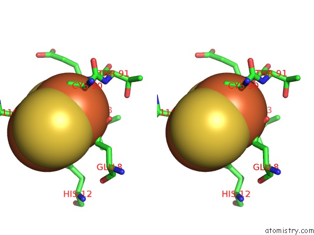
Iron binding site 1 out of 4 in 6y42
Go back to
Iron binding site 1 out
of 4 in the Crystal Structure of Rsrr Complexed to A 39 Basepair Dna Fragment of the Rsrr Promoter

Mono view

Stereo pair view
Mono view
Stereo pair view
A full contact list of Iron with other atoms in the Fe binding
site number 1 of Crystal Structure of Rsrr Complexed to A 39 Basepair Dna Fragment of the Rsrr Promoter within 5.0Å range:
|
Iron binding site 2 out of 4 in 6y42
Go back to
Iron binding site 2 out
of 4 in the Crystal Structure of Rsrr Complexed to A 39 Basepair Dna Fragment of the Rsrr Promoter

Mono view

Stereo pair view
Mono view
Stereo pair view
A full contact list of Iron with other atoms in the Fe binding
site number 2 of Crystal Structure of Rsrr Complexed to A 39 Basepair Dna Fragment of the Rsrr Promoter within 5.0Å range:
|
Iron binding site 3 out of 4 in 6y42
Go back to
Iron binding site 3 out
of 4 in the Crystal Structure of Rsrr Complexed to A 39 Basepair Dna Fragment of the Rsrr Promoter

Mono view
Stereo pair view
Mono view
Stereo pair view
A full contact list of Iron with other atoms in the Fe binding
site number 3 of Crystal Structure of Rsrr Complexed to A 39 Basepair Dna Fragment of the Rsrr Promoter within 5.0Å range:
|
Iron binding site 4 out of 4 in 6y42
Go back to
Iron binding site 4 out
of 4 in the Crystal Structure of Rsrr Complexed to A 39 Basepair Dna Fragment of the Rsrr Promoter

Mono view

Stereo pair view
Mono view
Stereo pair view
A full contact list of Iron with other atoms in the Fe binding
site number 4 of Crystal Structure of Rsrr Complexed to A 39 Basepair Dna Fragment of the Rsrr Promoter within 5.0Å range:
|
Reference:
J.C.Crack,
P.Amara,
A.Volbeda,
J.M.Mouesca,
R.Rohac,
M.T.Pellicer Martinez,
C.Y.Huang,
O.Gigarel,
C.Rinaldi,
N.E.Le Brun,
J.C.Fontecilla-Camps.
Electron and Proton Transfers Modulate Dna Binding By the Transcription Regulator Rsrr. J.Am.Chem.Soc. 2020.
ISSN: ESSN 1520-5126
PubMed: 32078310
DOI: 10.1021/JACS.9B12250
Page generated: Wed Aug 7 15:50:37 2024
ISSN: ESSN 1520-5126
PubMed: 32078310
DOI: 10.1021/JACS.9B12250
Last articles
Fe in 2YXOFe in 2YRS
Fe in 2YXC
Fe in 2YNM
Fe in 2YVJ
Fe in 2YP1
Fe in 2YU2
Fe in 2YU1
Fe in 2YQB
Fe in 2YOO