Iron »
PDB 6xz8-6yf4 »
6y8s »
Iron in PDB 6y8s: Crystal Structure of the Quaternary Ammonium Rieske Monooxygenase Cnta in Complex with Substrate Gamma-Butyrobetaine
Enzymatic activity of Crystal Structure of the Quaternary Ammonium Rieske Monooxygenase Cnta in Complex with Substrate Gamma-Butyrobetaine
All present enzymatic activity of Crystal Structure of the Quaternary Ammonium Rieske Monooxygenase Cnta in Complex with Substrate Gamma-Butyrobetaine:
1.14.13.239;
1.14.13.239;
Protein crystallography data
The structure of Crystal Structure of the Quaternary Ammonium Rieske Monooxygenase Cnta in Complex with Substrate Gamma-Butyrobetaine, PDB code: 6y8s
was solved by
M.Quareshy,
M.Shanmugam,
T.D.Bugg,
A.Cameron,
Y.Chen,
with X-Ray Crystallography technique. A brief refinement statistics is given in the table below:
| Resolution Low / High (Å) | 39.78 / 1.63 |
| Space group | P 63 |
| Cell size a, b, c (Å), α, β, γ (°) | 91.160, 91.160, 81.470, 90.00, 90.00, 120.00 |
| R / Rfree (%) | 17 / 19.3 |
Iron Binding Sites:
The binding sites of Iron atom in the Crystal Structure of the Quaternary Ammonium Rieske Monooxygenase Cnta in Complex with Substrate Gamma-Butyrobetaine
(pdb code 6y8s). This binding sites where shown within
5.0 Angstroms radius around Iron atom.
In total 3 binding sites of Iron where determined in the Crystal Structure of the Quaternary Ammonium Rieske Monooxygenase Cnta in Complex with Substrate Gamma-Butyrobetaine, PDB code: 6y8s:
Jump to Iron binding site number: 1; 2; 3;
In total 3 binding sites of Iron where determined in the Crystal Structure of the Quaternary Ammonium Rieske Monooxygenase Cnta in Complex with Substrate Gamma-Butyrobetaine, PDB code: 6y8s:
Jump to Iron binding site number: 1; 2; 3;
Iron binding site 1 out of 3 in 6y8s
Go back to
Iron binding site 1 out
of 3 in the Crystal Structure of the Quaternary Ammonium Rieske Monooxygenase Cnta in Complex with Substrate Gamma-Butyrobetaine
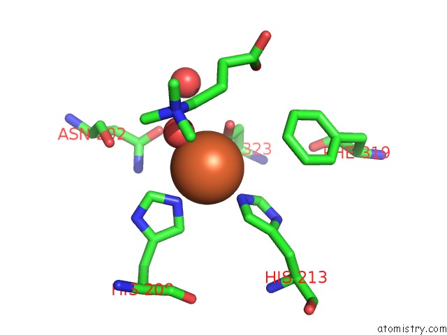
Mono view
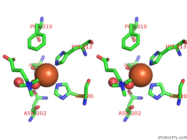
Stereo pair view
Mono view
Stereo pair view
A full contact list of Iron with other atoms in the Fe binding
site number 1 of Crystal Structure of the Quaternary Ammonium Rieske Monooxygenase Cnta in Complex with Substrate Gamma-Butyrobetaine within 5.0Å range:
|
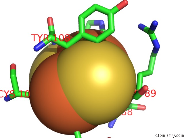
Iron binding site 2 out of 3 in 6y8s
Go back to
Iron binding site 2 out
of 3 in the Crystal Structure of the Quaternary Ammonium Rieske Monooxygenase Cnta in Complex with Substrate Gamma-Butyrobetaine
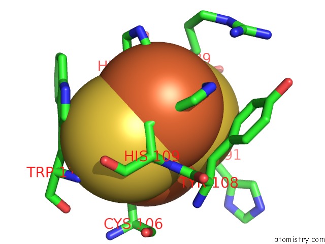
Mono view

Stereo pair view
Mono view
Stereo pair view
A full contact list of Iron with other atoms in the Fe binding
site number 2 of Crystal Structure of the Quaternary Ammonium Rieske Monooxygenase Cnta in Complex with Substrate Gamma-Butyrobetaine within 5.0Å range:
|
Iron binding site 3 out of 3 in 6y8s
Go back to
Iron binding site 3 out
of 3 in the Crystal Structure of the Quaternary Ammonium Rieske Monooxygenase Cnta in Complex with Substrate Gamma-Butyrobetaine
Mono view

Stereo pair view
Mono view
Stereo pair view
A full contact list of Iron with other atoms in the Fe binding
site number 3 of Crystal Structure of the Quaternary Ammonium Rieske Monooxygenase Cnta in Complex with Substrate Gamma-Butyrobetaine within 5.0Å range:
|
Reference:
M.Quareshy,
M.Shanmugam,
E.Townsend,
E.Jameson,
T.D.H.Bugg,
A.D.Cameron,
Y.Chen.
Structural Basis of Carnitine Monooxygenase Cnta Substrate Specificity, Inhibition and Inter-Subunit Electron Transfer. J.Biol.Chem. 2020.
ISSN: ESSN 1083-351X
PubMed: 33158989
DOI: 10.1074/JBC.RA120.016019
Page generated: Wed Aug 7 15:53:42 2024
ISSN: ESSN 1083-351X
PubMed: 33158989
DOI: 10.1074/JBC.RA120.016019
Last articles
Zn in 9MJ5Zn in 9HNW
Zn in 9G0L
Zn in 9FNE
Zn in 9DZN
Zn in 9E0I
Zn in 9D32
Zn in 9DAK
Zn in 8ZXC
Zn in 8ZUF