Iron »
PDB 6zzx-7ah6 »
7a4l »
Iron in PDB 7a4l: Pre-Only Solution Structure of the Iron-Sulfur Protein Pioc From Rhodopseudomonas Palustris Tie-1
Iron Binding Sites:
The binding sites of Iron atom in the Pre-Only Solution Structure of the Iron-Sulfur Protein Pioc From Rhodopseudomonas Palustris Tie-1
(pdb code 7a4l). This binding sites where shown within
5.0 Angstroms radius around Iron atom.
In total 4 binding sites of Iron where determined in the Pre-Only Solution Structure of the Iron-Sulfur Protein Pioc From Rhodopseudomonas Palustris Tie-1, PDB code: 7a4l:
Jump to Iron binding site number: 1; 2; 3; 4;
In total 4 binding sites of Iron where determined in the Pre-Only Solution Structure of the Iron-Sulfur Protein Pioc From Rhodopseudomonas Palustris Tie-1, PDB code: 7a4l:
Jump to Iron binding site number: 1; 2; 3; 4;
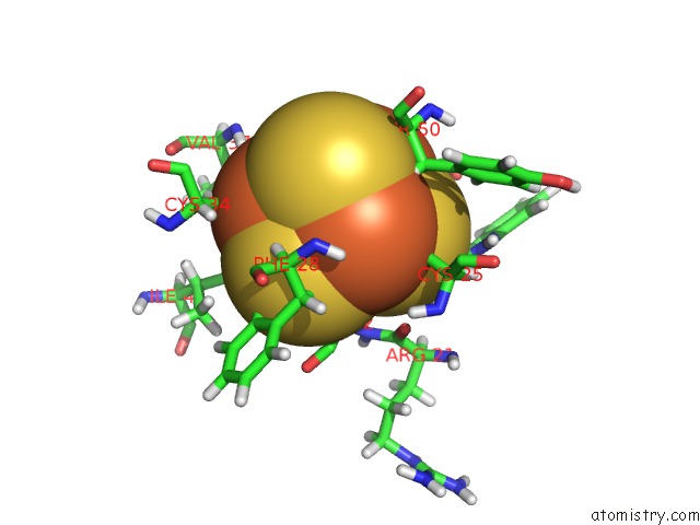
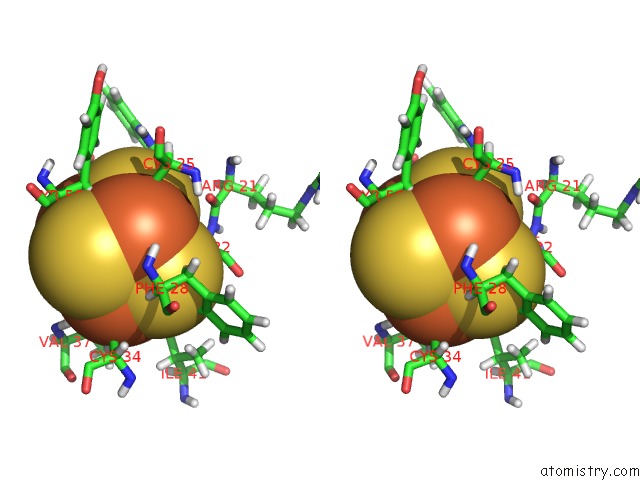
Iron binding site 1 out of 4 in 7a4l
Go back to
Iron binding site 1 out
of 4 in the Pre-Only Solution Structure of the Iron-Sulfur Protein Pioc From Rhodopseudomonas Palustris Tie-1
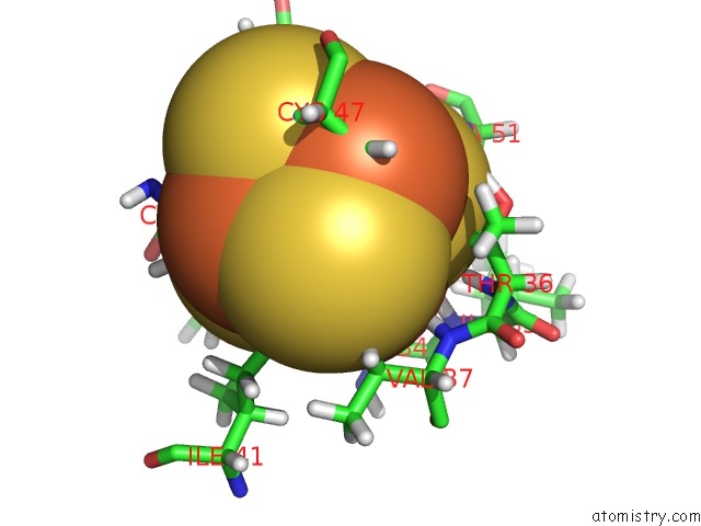
Mono view
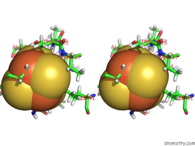
Stereo pair view
Mono view
Stereo pair view
A full contact list of Iron with other atoms in the Fe binding
site number 1 of Pre-Only Solution Structure of the Iron-Sulfur Protein Pioc From Rhodopseudomonas Palustris Tie-1 within 5.0Å range:
|
Iron binding site 2 out of 4 in 7a4l
Go back to
Iron binding site 2 out
of 4 in the Pre-Only Solution Structure of the Iron-Sulfur Protein Pioc From Rhodopseudomonas Palustris Tie-1
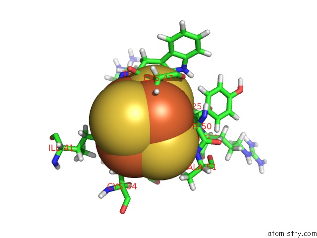
Mono view
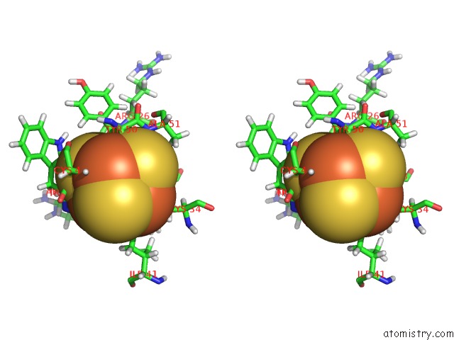
Stereo pair view
Mono view
Stereo pair view
A full contact list of Iron with other atoms in the Fe binding
site number 2 of Pre-Only Solution Structure of the Iron-Sulfur Protein Pioc From Rhodopseudomonas Palustris Tie-1 within 5.0Å range:
|
Iron binding site 3 out of 4 in 7a4l
Go back to
Iron binding site 3 out
of 4 in the Pre-Only Solution Structure of the Iron-Sulfur Protein Pioc From Rhodopseudomonas Palustris Tie-1
Mono view
Stereo pair view
Mono view
Stereo pair view
A full contact list of Iron with other atoms in the Fe binding
site number 3 of Pre-Only Solution Structure of the Iron-Sulfur Protein Pioc From Rhodopseudomonas Palustris Tie-1 within 5.0Å range:
|
Iron binding site 4 out of 4 in 7a4l
Go back to
Iron binding site 4 out
of 4 in the Pre-Only Solution Structure of the Iron-Sulfur Protein Pioc From Rhodopseudomonas Palustris Tie-1
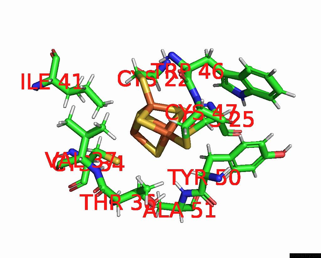
Mono view
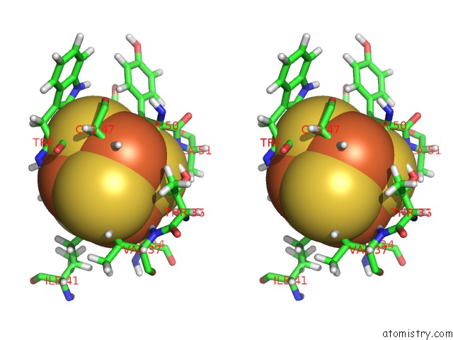
Stereo pair view
Mono view
Stereo pair view
A full contact list of Iron with other atoms in the Fe binding
site number 4 of Pre-Only Solution Structure of the Iron-Sulfur Protein Pioc From Rhodopseudomonas Palustris Tie-1 within 5.0Å range:
|
Reference:
I.B.Trindade,
M.Invernici,
F.Cantini,
R.O.Louro,
M.Piccioli.
Pre-Driven Protein uc(Nmr) Structures: An Alternative Approach in Highly Paramagnetic Systems. Febs J. 2020.
ISSN: ISSN 1742-464X
PubMed: 33124176
DOI: 10.1111/FEBS.15615
Page generated: Wed Aug 7 21:43:27 2024
ISSN: ISSN 1742-464X
PubMed: 33124176
DOI: 10.1111/FEBS.15615
Last articles
Zn in 9J0NZn in 9J0O
Zn in 9J0P
Zn in 9FJX
Zn in 9EKB
Zn in 9C0F
Zn in 9CAH
Zn in 9CH0
Zn in 9CH3
Zn in 9CH1