Iron »
PDB 7ai8-7bha »
7ai8 »
Iron in PDB 7ai8: Structure of Ribonucleotide Reductase R2 From Escherichia Coli Collected By Still Serial Crystallography on A Coc Membrane at A Synchrotron Source
Enzymatic activity of Structure of Ribonucleotide Reductase R2 From Escherichia Coli Collected By Still Serial Crystallography on A Coc Membrane at A Synchrotron Source
All present enzymatic activity of Structure of Ribonucleotide Reductase R2 From Escherichia Coli Collected By Still Serial Crystallography on A Coc Membrane at A Synchrotron Source:
1.17.4.1;
1.17.4.1;
Protein crystallography data
The structure of Structure of Ribonucleotide Reductase R2 From Escherichia Coli Collected By Still Serial Crystallography on A Coc Membrane at A Synchrotron Source, PDB code: 7ai8
was solved by
O.Aurelius,
J.John,
I.Martiel,
C.Padeste,
A.Karpik,
C.Y.Huang,
M.Hogbom,
M.Wang,
M.Marsh,
with X-Ray Crystallography technique. A brief refinement statistics is given in the table below:
| Resolution Low / High (Å) | 51.97 / 2.10 |
| Space group | P 61 2 2 |
| Cell size a, b, c (Å), α, β, γ (°) | 90.35, 90.35, 208.55, 90, 90, 120 |
| R / Rfree (%) | 19 / 21.9 |
Iron Binding Sites:
The binding sites of Iron atom in the Structure of Ribonucleotide Reductase R2 From Escherichia Coli Collected By Still Serial Crystallography on A Coc Membrane at A Synchrotron Source
(pdb code 7ai8). This binding sites where shown within
5.0 Angstroms radius around Iron atom.
In total 2 binding sites of Iron where determined in the Structure of Ribonucleotide Reductase R2 From Escherichia Coli Collected By Still Serial Crystallography on A Coc Membrane at A Synchrotron Source, PDB code: 7ai8:
Jump to Iron binding site number: 1; 2;
In total 2 binding sites of Iron where determined in the Structure of Ribonucleotide Reductase R2 From Escherichia Coli Collected By Still Serial Crystallography on A Coc Membrane at A Synchrotron Source, PDB code: 7ai8:
Jump to Iron binding site number: 1; 2;
Iron binding site 1 out of 2 in 7ai8
Go back to
Iron binding site 1 out
of 2 in the Structure of Ribonucleotide Reductase R2 From Escherichia Coli Collected By Still Serial Crystallography on A Coc Membrane at A Synchrotron Source
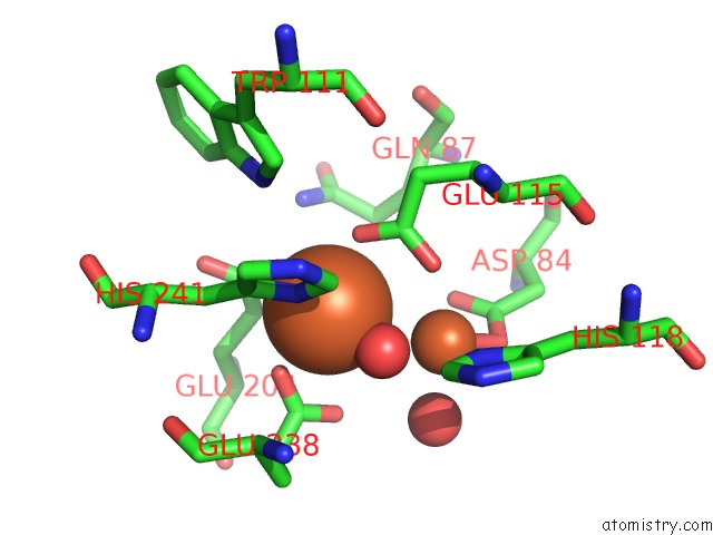
Mono view
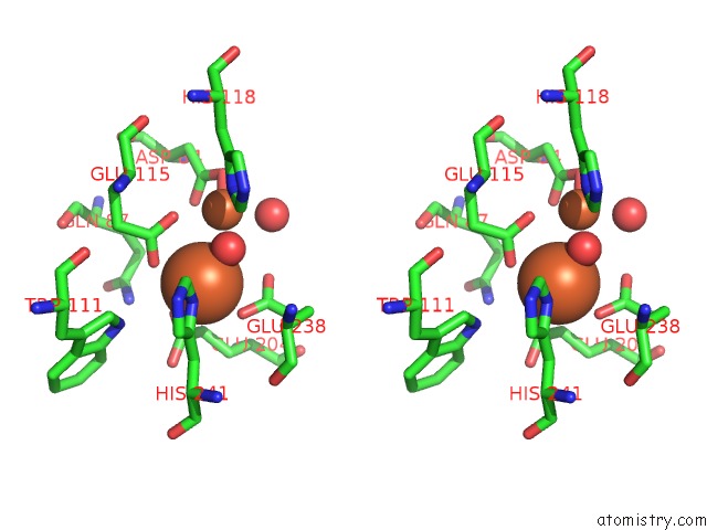
Stereo pair view
Mono view
Stereo pair view
A full contact list of Iron with other atoms in the Fe binding
site number 1 of Structure of Ribonucleotide Reductase R2 From Escherichia Coli Collected By Still Serial Crystallography on A Coc Membrane at A Synchrotron Source within 5.0Å range:
|
Iron binding site 2 out of 2 in 7ai8
Go back to
Iron binding site 2 out
of 2 in the Structure of Ribonucleotide Reductase R2 From Escherichia Coli Collected By Still Serial Crystallography on A Coc Membrane at A Synchrotron Source
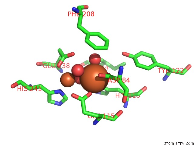
Mono view
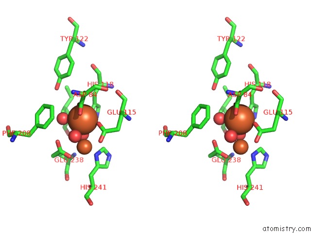
Stereo pair view
Mono view
Stereo pair view
A full contact list of Iron with other atoms in the Fe binding
site number 2 of Structure of Ribonucleotide Reductase R2 From Escherichia Coli Collected By Still Serial Crystallography on A Coc Membrane at A Synchrotron Source within 5.0Å range:
|
Reference:
I.Martiel,
J.H.Beale,
A.Karpik,
C.Y.Huang,
L.Vera,
N.Olieric,
M.Wranik,
C.J.Tsai,
J.Muhle,
O.Aurelius,
J.John,
M.Hogbom,
M.Wang,
M.Marsh,
C.Padeste.
Versatile Microporous Polymer-Based Supports For Serial Macromolecular Crystallography Acta Crystallogr.,Sect.D 2021.
ISSN: ESSN 1399-0047
DOI: 10.1107/S2059798321007324
Page generated: Wed Aug 7 22:30:21 2024
ISSN: ESSN 1399-0047
DOI: 10.1107/S2059798321007324
Last articles
Zn in 9J0NZn in 9J0O
Zn in 9J0P
Zn in 9FJX
Zn in 9EKB
Zn in 9C0F
Zn in 9CAH
Zn in 9CH0
Zn in 9CH3
Zn in 9CH1