Iron »
PDB 7ai8-7bha »
7ant »
Iron in PDB 7ant: Structure of CYP153A From Polaromonas Sp.
Protein crystallography data
The structure of Structure of CYP153A From Polaromonas Sp., PDB code: 7ant
was solved by
E.Zukic,
B.Rowlinson,
M.Sharma,
S.Hoffman,
B.Hauer,
G.Grogan,
with X-Ray Crystallography technique. A brief refinement statistics is given in the table below:
| Resolution Low / High (Å) | 58.64 / 1.52 |
| Space group | P 21 21 21 |
| Cell size a, b, c (Å), α, β, γ (°) | 72.852, 98.501, 112.627, 90, 90, 90 |
| R / Rfree (%) | 20.7 / 23.1 |
Iron Binding Sites:
The binding sites of Iron atom in the Structure of CYP153A From Polaromonas Sp.
(pdb code 7ant). This binding sites where shown within
5.0 Angstroms radius around Iron atom.
In total 2 binding sites of Iron where determined in the Structure of CYP153A From Polaromonas Sp., PDB code: 7ant:
Jump to Iron binding site number: 1; 2;
In total 2 binding sites of Iron where determined in the Structure of CYP153A From Polaromonas Sp., PDB code: 7ant:
Jump to Iron binding site number: 1; 2;
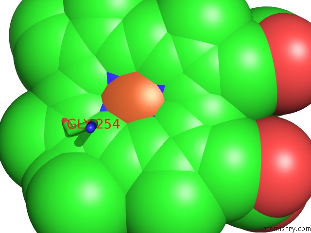
Iron binding site 1 out of 2 in 7ant
Go back to
Iron binding site 1 out
of 2 in the Structure of CYP153A From Polaromonas Sp.

Mono view

Stereo pair view
Mono view
Stereo pair view
A full contact list of Iron with other atoms in the Fe binding
site number 1 of Structure of CYP153A From Polaromonas Sp. within 5.0Å range:
|
Iron binding site 2 out of 2 in 7ant
Go back to
Iron binding site 2 out
of 2 in the Structure of CYP153A From Polaromonas Sp.
Mono view

Stereo pair view
Mono view
Stereo pair view
A full contact list of Iron with other atoms in the Fe binding
site number 2 of Structure of CYP153A From Polaromonas Sp. within 5.0Å range:
|
Reference:
L.R.Rapp,
S.M.Marques,
E.Zukic,
B.Rowlinson,
M.Sharma,
G.Grogan,
J.Damborsky,
B.Hauer.
Substrate Anchoring and Flexibility Reduction in CYP153AM.Aq Leads to Highly Improved Efficiency Toward Octanoic Acid Acs Catalysis V. 11 3182 2021.
ISSN: ESSN 2155-5435
DOI: 10.1021/ACSCATAL.0C05193
Page generated: Wed Aug 7 22:30:21 2024
ISSN: ESSN 2155-5435
DOI: 10.1021/ACSCATAL.0C05193
Last articles
F in 4JTQF in 4JSC
F in 4JSJ
F in 4JSM
F in 4JQG
F in 4JSI
F in 4JQ2
F in 4JP4
F in 4JPS
F in 4JNC