Iron »
PDB 7bhb-7cji »
7c8n »
Iron in PDB 7c8n: Crystal Structure of Iscu H106A Variant
Protein crystallography data
The structure of Crystal Structure of Iscu H106A Variant, PDB code: 7c8n
was solved by
K.Kunichika,
Y.Takahashi,
T.Fujishiro,
with X-Ray Crystallography technique. A brief refinement statistics is given in the table below:
| Resolution Low / High (Å) | 41.54 / 1.50 |
| Space group | P 63 2 2 |
| Cell size a, b, c (Å), α, β, γ (°) | 107.021, 107.021, 65.753, 90, 90, 120 |
| R / Rfree (%) | 17.7 / 19.7 |
Iron Binding Sites:
The binding sites of Iron atom in the Crystal Structure of Iscu H106A Variant
(pdb code 7c8n). This binding sites where shown within
5.0 Angstroms radius around Iron atom.
In total 2 binding sites of Iron where determined in the Crystal Structure of Iscu H106A Variant, PDB code: 7c8n:
Jump to Iron binding site number: 1; 2;
In total 2 binding sites of Iron where determined in the Crystal Structure of Iscu H106A Variant, PDB code: 7c8n:
Jump to Iron binding site number: 1; 2;
Iron binding site 1 out of 2 in 7c8n
Go back to
Iron binding site 1 out
of 2 in the Crystal Structure of Iscu H106A Variant
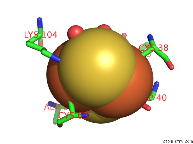
Mono view
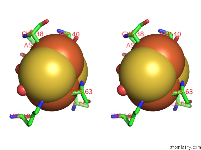
Stereo pair view
Mono view
Stereo pair view
A full contact list of Iron with other atoms in the Fe binding
site number 1 of Crystal Structure of Iscu H106A Variant within 5.0Å range:
|
Iron binding site 2 out of 2 in 7c8n
Go back to
Iron binding site 2 out
of 2 in the Crystal Structure of Iscu H106A Variant
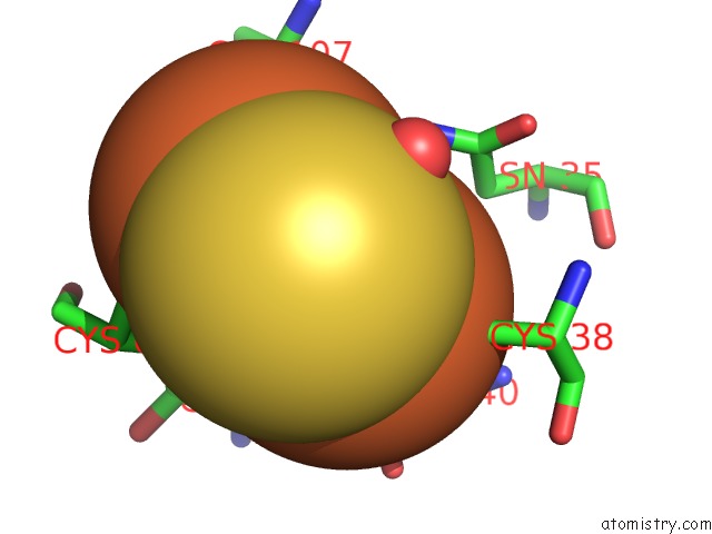
Mono view
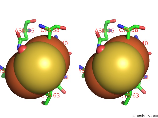
Stereo pair view
Mono view
Stereo pair view
A full contact list of Iron with other atoms in the Fe binding
site number 2 of Crystal Structure of Iscu H106A Variant within 5.0Å range:
|
Reference:
K.Kunichika,
R.Nakamura,
T.Fujishiro,
Y.Takahashi.
The Structure of the Dimeric State of Iscu Harboring Two Adjacent [2FE-2S] Clusters Provides Mechanistic Insights Into Cluster Conversion to [4FE-4S]. Biochemistry 2021.
ISSN: ISSN 0006-2960
PubMed: 33938220
DOI: 10.1021/ACS.BIOCHEM.1C00112
Page generated: Thu Aug 8 00:35:30 2024
ISSN: ISSN 0006-2960
PubMed: 33938220
DOI: 10.1021/ACS.BIOCHEM.1C00112
Last articles
Zn in 9J0NZn in 9J0O
Zn in 9J0P
Zn in 9FJX
Zn in 9EKB
Zn in 9C0F
Zn in 9CAH
Zn in 9CH0
Zn in 9CH3
Zn in 9CH1