Iron »
PDB 7cjj-7czi »
7coh »
Iron in PDB 7coh: Dimeric Form of Bovine Heart Cytochrome C Oxidase in the Fully Oxidized State
Enzymatic activity of Dimeric Form of Bovine Heart Cytochrome C Oxidase in the Fully Oxidized State
All present enzymatic activity of Dimeric Form of Bovine Heart Cytochrome C Oxidase in the Fully Oxidized State:
7.1.1.9;
7.1.1.9;
Protein crystallography data
The structure of Dimeric Form of Bovine Heart Cytochrome C Oxidase in the Fully Oxidized State, PDB code: 7coh
was solved by
K.Shinzawa-Itoh,
K.Muramoto,
with X-Ray Crystallography technique. A brief refinement statistics is given in the table below:
| Resolution Low / High (Å) | 40.00 / 1.30 |
| Space group | P 21 21 21 |
| Cell size a, b, c (Å), α, β, γ (°) | 181.998, 204.193, 177.759, 90, 90, 90 |
| R / Rfree (%) | 14.9 / 17 |
Other elements in 7coh:
The structure of Dimeric Form of Bovine Heart Cytochrome C Oxidase in the Fully Oxidized State also contains other interesting chemical elements:
| Magnesium | (Mg) | 2 atoms |
| Zinc | (Zn) | 2 atoms |
| Copper | (Cu) | 6 atoms |
| Sodium | (Na) | 2 atoms |
Iron Binding Sites:
The binding sites of Iron atom in the Dimeric Form of Bovine Heart Cytochrome C Oxidase in the Fully Oxidized State
(pdb code 7coh). This binding sites where shown within
5.0 Angstroms radius around Iron atom.
In total 4 binding sites of Iron where determined in the Dimeric Form of Bovine Heart Cytochrome C Oxidase in the Fully Oxidized State, PDB code: 7coh:
Jump to Iron binding site number: 1; 2; 3; 4;
In total 4 binding sites of Iron where determined in the Dimeric Form of Bovine Heart Cytochrome C Oxidase in the Fully Oxidized State, PDB code: 7coh:
Jump to Iron binding site number: 1; 2; 3; 4;
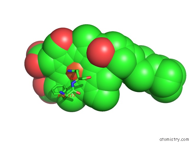
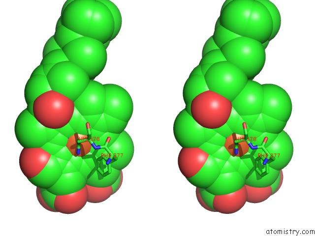
Iron binding site 1 out of 4 in 7coh
Go back to
Iron binding site 1 out
of 4 in the Dimeric Form of Bovine Heart Cytochrome C Oxidase in the Fully Oxidized State
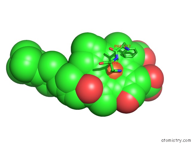
Mono view
Stereo pair view
Mono view
Stereo pair view
A full contact list of Iron with other atoms in the Fe binding
site number 1 of Dimeric Form of Bovine Heart Cytochrome C Oxidase in the Fully Oxidized State within 5.0Å range:
|
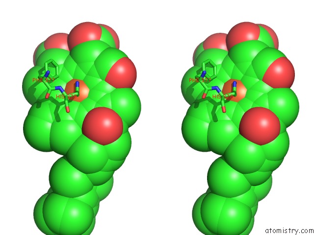
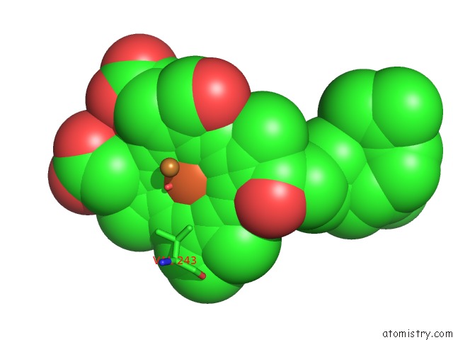
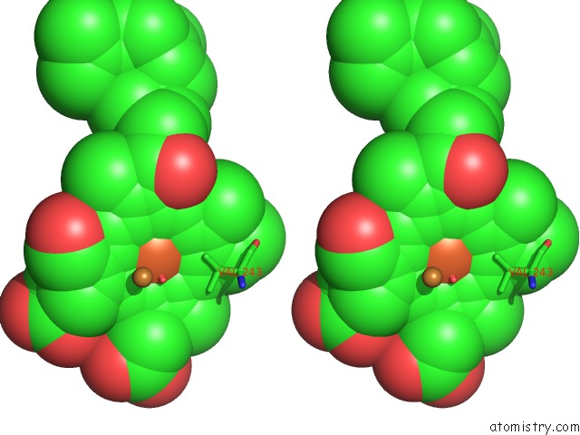
Iron binding site 2 out of 4 in 7coh
Go back to
Iron binding site 2 out
of 4 in the Dimeric Form of Bovine Heart Cytochrome C Oxidase in the Fully Oxidized State
Mono view
Stereo pair view
Mono view
Stereo pair view
A full contact list of Iron with other atoms in the Fe binding
site number 2 of Dimeric Form of Bovine Heart Cytochrome C Oxidase in the Fully Oxidized State within 5.0Å range:
|
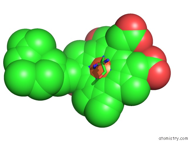
Iron binding site 3 out of 4 in 7coh
Go back to
Iron binding site 3 out
of 4 in the Dimeric Form of Bovine Heart Cytochrome C Oxidase in the Fully Oxidized State
Mono view
Stereo pair view
Mono view
Stereo pair view
A full contact list of Iron with other atoms in the Fe binding
site number 3 of Dimeric Form of Bovine Heart Cytochrome C Oxidase in the Fully Oxidized State within 5.0Å range:
|
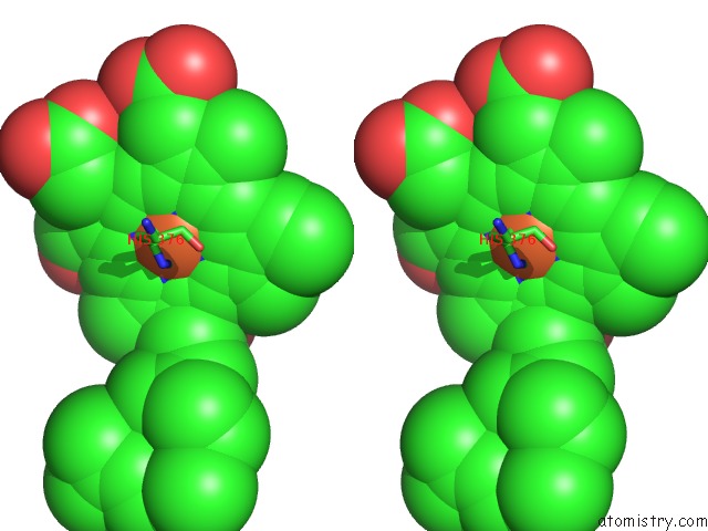
Iron binding site 4 out of 4 in 7coh
Go back to
Iron binding site 4 out
of 4 in the Dimeric Form of Bovine Heart Cytochrome C Oxidase in the Fully Oxidized State
Mono view
Stereo pair view
Mono view
Stereo pair view
A full contact list of Iron with other atoms in the Fe binding
site number 4 of Dimeric Form of Bovine Heart Cytochrome C Oxidase in the Fully Oxidized State within 5.0Å range:
|
Reference:
K.Shinzawa-Itoh,
M.Hatanaka,
K.Fujita,
N.Yano,
Y.Ogasawara,
J.Iwata,
E.Yamashita,
T.Tsukihara,
S.Yoshikawa,
K.Muramoto.
The 1.3-A Resolution Structure of Bovine Cytochrome C Oxidase Suggests A Dimerization Mechanism Biochim.Biophys.Acta 2021.
ISSN: ISSN 0006-3002
DOI: 10.1016/J.BBADVA.2021.100009
Page generated: Thu Aug 8 03:11:53 2024
ISSN: ISSN 0006-3002
DOI: 10.1016/J.BBADVA.2021.100009
Last articles
Zn in 9J0NZn in 9J0O
Zn in 9J0P
Zn in 9FJX
Zn in 9EKB
Zn in 9C0F
Zn in 9CAH
Zn in 9CH0
Zn in 9CH3
Zn in 9CH1