Iron »
PDB 7dgl-7dya »
7dgm »
Iron in PDB 7dgm: The Dimeric Structure of K79H/G80A/H81A Myoglobin
Protein crystallography data
The structure of The Dimeric Structure of K79H/G80A/H81A Myoglobin, PDB code: 7dgm
was solved by
S.Nagao,
A.Idomoto,
N.Shibata,
Y.Higuchi,
S.Hirota,
with X-Ray Crystallography technique. A brief refinement statistics is given in the table below:
| Resolution Low / High (Å) | 47.15 / 1.62 |
| Space group | P 21 21 21 |
| Cell size a, b, c (Å), α, β, γ (°) | 57.227, 62.897, 82.962, 90, 90, 90 |
| R / Rfree (%) | 20.2 / 22.3 |
Iron Binding Sites:
The binding sites of Iron atom in the The Dimeric Structure of K79H/G80A/H81A Myoglobin
(pdb code 7dgm). This binding sites where shown within
5.0 Angstroms radius around Iron atom.
In total 2 binding sites of Iron where determined in the The Dimeric Structure of K79H/G80A/H81A Myoglobin, PDB code: 7dgm:
Jump to Iron binding site number: 1; 2;
In total 2 binding sites of Iron where determined in the The Dimeric Structure of K79H/G80A/H81A Myoglobin, PDB code: 7dgm:
Jump to Iron binding site number: 1; 2;
Iron binding site 1 out of 2 in 7dgm
Go back to
Iron binding site 1 out
of 2 in the The Dimeric Structure of K79H/G80A/H81A Myoglobin
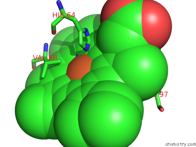
Mono view
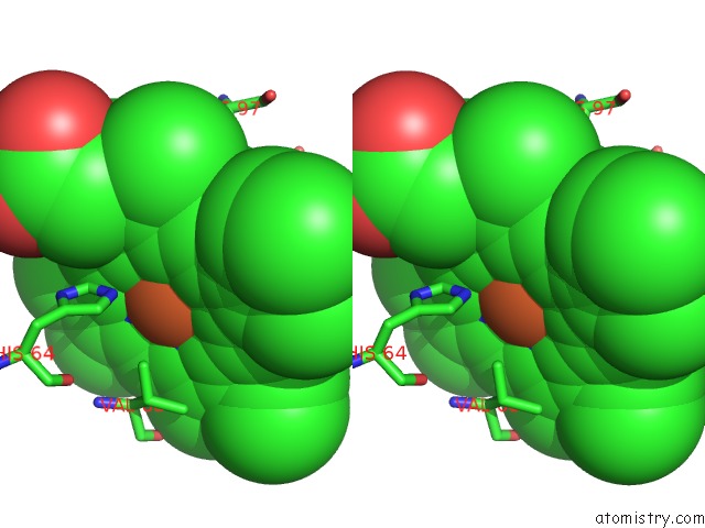
Stereo pair view
Mono view
Stereo pair view
A full contact list of Iron with other atoms in the Fe binding
site number 1 of The Dimeric Structure of K79H/G80A/H81A Myoglobin within 5.0Å range:
|
Iron binding site 2 out of 2 in 7dgm
Go back to
Iron binding site 2 out
of 2 in the The Dimeric Structure of K79H/G80A/H81A Myoglobin
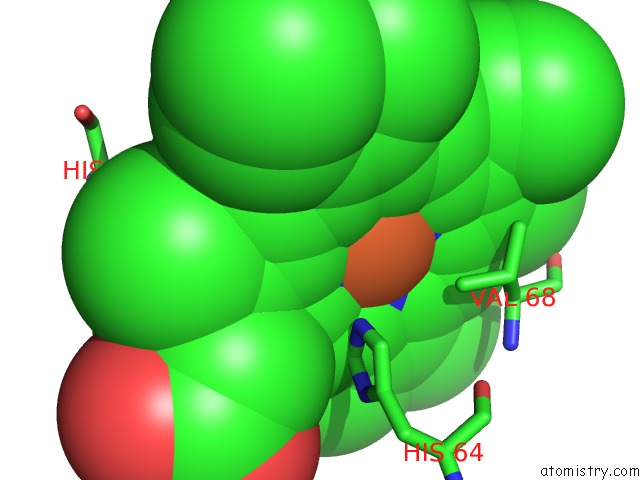
Mono view
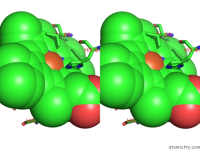
Stereo pair view
Mono view
Stereo pair view
A full contact list of Iron with other atoms in the Fe binding
site number 2 of The Dimeric Structure of K79H/G80A/H81A Myoglobin within 5.0Å range:
|
Reference:
S.Nagao,
A.Idomoto,
N.Shibata,
Y.Higuchi,
S.Hirota.
Rational Design of Metal-Binding Sites in Domain-Swapped Myoglobin Dimers. J.Inorg.Biochem. 2021.
ISSN: ISSN 0162-0134
DOI: 10.1016/J.JINORGBIO.2021.111374
Page generated: Wed Aug 6 21:39:47 2025
ISSN: ISSN 0162-0134
DOI: 10.1016/J.JINORGBIO.2021.111374
Last articles
Fe in 8RNRFe in 8RIU
Fe in 8RNQ
Fe in 8RNP
Fe in 8RNN
Fe in 8RNM
Fe in 8RNL
Fe in 8RNJ
Fe in 8RNK
Fe in 8RMG