Iron »
PDB 7dgk-7dy8 »
7dvt »
Iron in PDB 7dvt: Crystal Structure of Heme Sensor Protein Pefr in Complex with Heme and Carbon Monoxide
Protein crystallography data
The structure of Crystal Structure of Heme Sensor Protein Pefr in Complex with Heme and Carbon Monoxide, PDB code: 7dvt
was solved by
M.Nishinaga,
S.Nagai,
Y.Nishitani,
H.Sugimoto,
Y.Shiro,
H.Sawai,
with X-Ray Crystallography technique. A brief refinement statistics is given in the table below:
| Resolution Low / High (Å) | 38.22 / 2.09 |
| Space group | C 1 2 1 |
| Cell size a, b, c (Å), α, β, γ (°) | 68.203, 31.331, 76.609, 90, 94.34, 90 |
| R / Rfree (%) | 22.6 / 28.7 |
Iron Binding Sites:
The binding sites of Iron atom in the Crystal Structure of Heme Sensor Protein Pefr in Complex with Heme and Carbon Monoxide
(pdb code 7dvt). This binding sites where shown within
5.0 Angstroms radius around Iron atom.
In total only one binding site of Iron was determined in the Crystal Structure of Heme Sensor Protein Pefr in Complex with Heme and Carbon Monoxide, PDB code: 7dvt:
In total only one binding site of Iron was determined in the Crystal Structure of Heme Sensor Protein Pefr in Complex with Heme and Carbon Monoxide, PDB code: 7dvt:
Iron binding site 1 out of 1 in 7dvt
Go back to
Iron binding site 1 out
of 1 in the Crystal Structure of Heme Sensor Protein Pefr in Complex with Heme and Carbon Monoxide
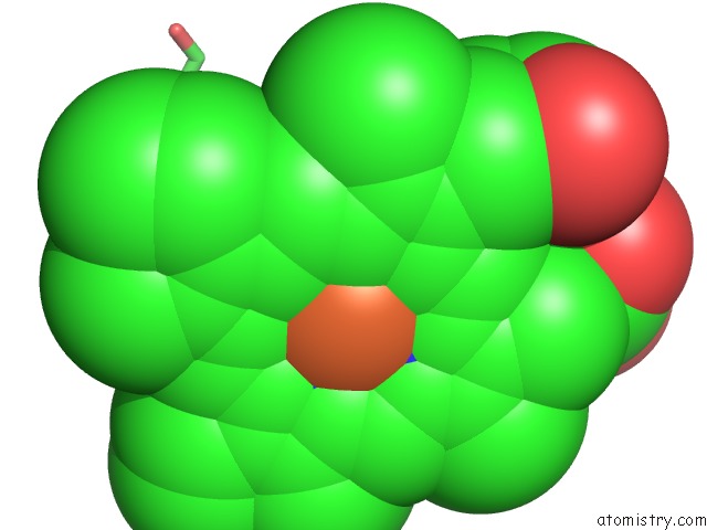
Mono view
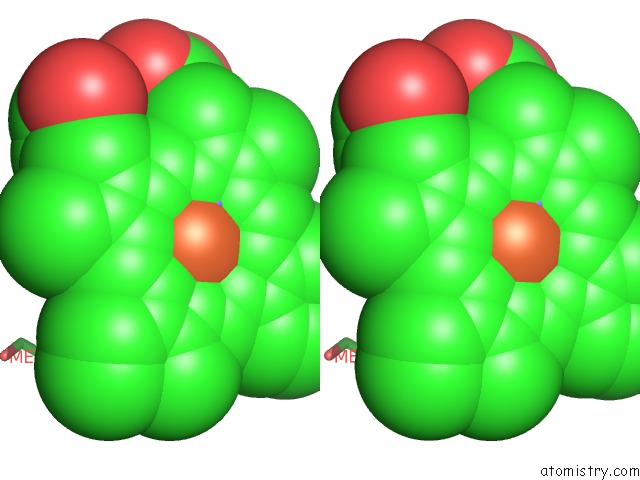
Stereo pair view
Mono view
Stereo pair view
A full contact list of Iron with other atoms in the Fe binding
site number 1 of Crystal Structure of Heme Sensor Protein Pefr in Complex with Heme and Carbon Monoxide within 5.0Å range:
|
Reference:
M.Nishinaga,
H.Sugimoto,
Y.Nishitani,
S.Nagai,
S.Nagatoishi,
N.Muraki,
T.Tosha,
K.Tsumoto,
S.Aono,
Y.Shiro,
H.Sawai.
Heme Controls the Structural Rearrangement of Its Sensor Protein Mediating the Hemolytic Bacterial Survival. Commun Biol V. 4 467 2021.
ISSN: ESSN 2399-3642
PubMed: 33850260
DOI: 10.1038/S42003-021-01987-5
Page generated: Thu Aug 8 04:19:07 2024
ISSN: ESSN 2399-3642
PubMed: 33850260
DOI: 10.1038/S42003-021-01987-5
Last articles
Zn in 9JYWZn in 9IR4
Zn in 9IR3
Zn in 9GMX
Zn in 9GMW
Zn in 9JEJ
Zn in 9ERF
Zn in 9ERE
Zn in 9EGV
Zn in 9EGW