Iron »
PDB 7dya-7ek4 »
7e5q »
Iron in PDB 7e5q: Crystal Structure of Dye Decolorizing Peroxidase From Bacillus Subtilis at Acidic pH
Protein crystallography data
The structure of Crystal Structure of Dye Decolorizing Peroxidase From Bacillus Subtilis at Acidic pH, PDB code: 7e5q
was solved by
P.Dhankhar,
V.Dalal,
P.Kumar,
with X-Ray Crystallography technique. A brief refinement statistics is given in the table below:
| Resolution Low / High (Å) | 44.34 / 1.90 |
| Space group | P 21 21 21 |
| Cell size a, b, c (Å), α, β, γ (°) | 40.855, 117.342, 135.441, 90, 90, 90 |
| R / Rfree (%) | 18 / 23.3 |
Other elements in 7e5q:
The structure of Crystal Structure of Dye Decolorizing Peroxidase From Bacillus Subtilis at Acidic pH also contains other interesting chemical elements:
| Sodium | (Na) | 8 atoms |
| Chlorine | (Cl) | 9 atoms |
Iron Binding Sites:
The binding sites of Iron atom in the Crystal Structure of Dye Decolorizing Peroxidase From Bacillus Subtilis at Acidic pH
(pdb code 7e5q). This binding sites where shown within
5.0 Angstroms radius around Iron atom.
In total 2 binding sites of Iron where determined in the Crystal Structure of Dye Decolorizing Peroxidase From Bacillus Subtilis at Acidic pH, PDB code: 7e5q:
Jump to Iron binding site number: 1; 2;
In total 2 binding sites of Iron where determined in the Crystal Structure of Dye Decolorizing Peroxidase From Bacillus Subtilis at Acidic pH, PDB code: 7e5q:
Jump to Iron binding site number: 1; 2;
Iron binding site 1 out of 2 in 7e5q
Go back to
Iron binding site 1 out
of 2 in the Crystal Structure of Dye Decolorizing Peroxidase From Bacillus Subtilis at Acidic pH
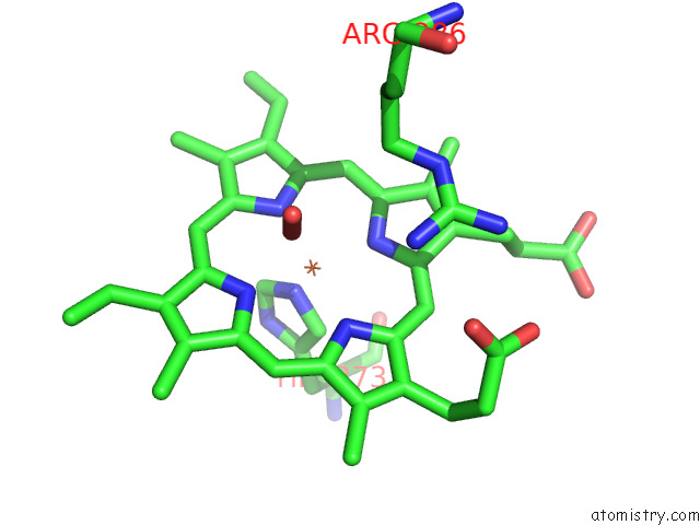
Mono view
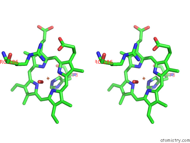
Stereo pair view
Mono view
Stereo pair view
A full contact list of Iron with other atoms in the Fe binding
site number 1 of Crystal Structure of Dye Decolorizing Peroxidase From Bacillus Subtilis at Acidic pH within 5.0Å range:
|
Iron binding site 2 out of 2 in 7e5q
Go back to
Iron binding site 2 out
of 2 in the Crystal Structure of Dye Decolorizing Peroxidase From Bacillus Subtilis at Acidic pH
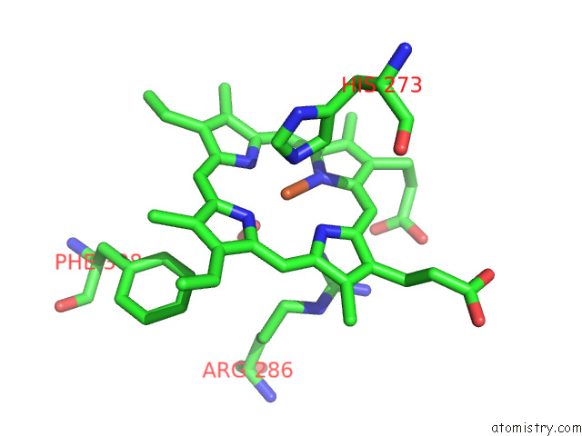
Mono view
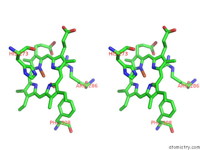
Stereo pair view
Mono view
Stereo pair view
A full contact list of Iron with other atoms in the Fe binding
site number 2 of Crystal Structure of Dye Decolorizing Peroxidase From Bacillus Subtilis at Acidic pH within 5.0Å range:
|
Reference:
P.Dhankhar,
V.Dalal,
A.K.Sharma,
P.Kumar.
Structural Insights at Acidic pH of Dye-Decolorizing Peroxidase From Bacillus Subtilis. Proteins 2022.
ISSN: ESSN 1097-0134
PubMed: 36345957
DOI: 10.1002/PROT.26444
Page generated: Thu Aug 8 04:56:38 2024
ISSN: ESSN 1097-0134
PubMed: 36345957
DOI: 10.1002/PROT.26444
Last articles
Zn in 9J0NZn in 9J0O
Zn in 9J0P
Zn in 9FJX
Zn in 9EKB
Zn in 9C0F
Zn in 9CAH
Zn in 9CH0
Zn in 9CH3
Zn in 9CH1