Iron »
PDB 7dya-7ek4 »
7egn »
Iron in PDB 7egn: Crystal Structure of the P450 BM3 Heme Domain Mutant F87A in Complex with N-Imidazolyl-Hexanoyl-L-Phenylalanine and Hydroxylamine
Enzymatic activity of Crystal Structure of the P450 BM3 Heme Domain Mutant F87A in Complex with N-Imidazolyl-Hexanoyl-L-Phenylalanine and Hydroxylamine
All present enzymatic activity of Crystal Structure of the P450 BM3 Heme Domain Mutant F87A in Complex with N-Imidazolyl-Hexanoyl-L-Phenylalanine and Hydroxylamine:
1.14.14.1; 1.6.2.4;
1.14.14.1; 1.6.2.4;
Protein crystallography data
The structure of Crystal Structure of the P450 BM3 Heme Domain Mutant F87A in Complex with N-Imidazolyl-Hexanoyl-L-Phenylalanine and Hydroxylamine, PDB code: 7egn
was solved by
Y.Jiang,
S.Dong,
Y.Feng,
Z.Cong,
with X-Ray Crystallography technique. A brief refinement statistics is given in the table below:
| Resolution Low / High (Å) | 39.43 / 2.70 |
| Space group | P 1 21 1 |
| Cell size a, b, c (Å), α, β, γ (°) | 58.67, 147.557, 64.324, 90, 99.98, 90 |
| R / Rfree (%) | 20 / 24.5 |
Iron Binding Sites:
The binding sites of Iron atom in the Crystal Structure of the P450 BM3 Heme Domain Mutant F87A in Complex with N-Imidazolyl-Hexanoyl-L-Phenylalanine and Hydroxylamine
(pdb code 7egn). This binding sites where shown within
5.0 Angstroms radius around Iron atom.
In total 2 binding sites of Iron where determined in the Crystal Structure of the P450 BM3 Heme Domain Mutant F87A in Complex with N-Imidazolyl-Hexanoyl-L-Phenylalanine and Hydroxylamine, PDB code: 7egn:
Jump to Iron binding site number: 1; 2;
In total 2 binding sites of Iron where determined in the Crystal Structure of the P450 BM3 Heme Domain Mutant F87A in Complex with N-Imidazolyl-Hexanoyl-L-Phenylalanine and Hydroxylamine, PDB code: 7egn:
Jump to Iron binding site number: 1; 2;
Iron binding site 1 out of 2 in 7egn
Go back to
Iron binding site 1 out
of 2 in the Crystal Structure of the P450 BM3 Heme Domain Mutant F87A in Complex with N-Imidazolyl-Hexanoyl-L-Phenylalanine and Hydroxylamine
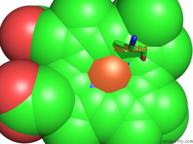
Mono view
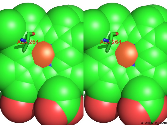
Stereo pair view
Mono view
Stereo pair view
A full contact list of Iron with other atoms in the Fe binding
site number 1 of Crystal Structure of the P450 BM3 Heme Domain Mutant F87A in Complex with N-Imidazolyl-Hexanoyl-L-Phenylalanine and Hydroxylamine within 5.0Å range:
|
Iron binding site 2 out of 2 in 7egn
Go back to
Iron binding site 2 out
of 2 in the Crystal Structure of the P450 BM3 Heme Domain Mutant F87A in Complex with N-Imidazolyl-Hexanoyl-L-Phenylalanine and Hydroxylamine
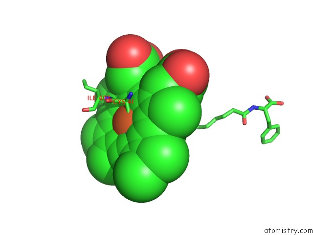
Mono view
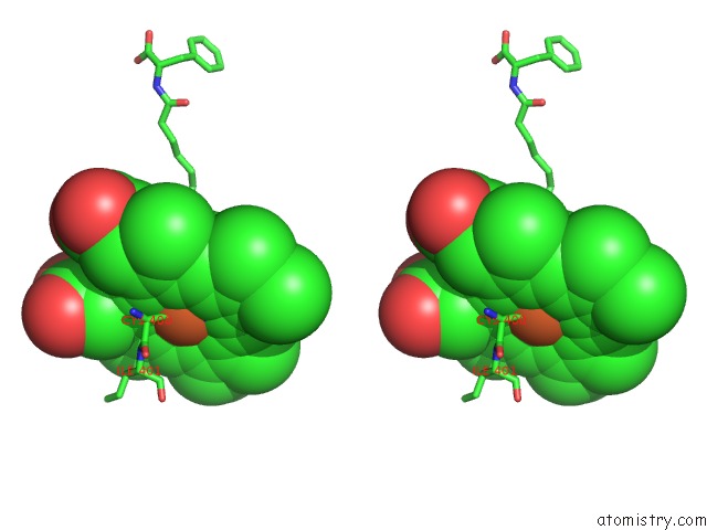
Stereo pair view
Mono view
Stereo pair view
A full contact list of Iron with other atoms in the Fe binding
site number 2 of Crystal Structure of the P450 BM3 Heme Domain Mutant F87A in Complex with N-Imidazolyl-Hexanoyl-L-Phenylalanine and Hydroxylamine within 5.0Å range:
|
Reference:
X.Zhang,
Y.Jiang,
Q.Chen,
S.Dong,
Y.Feng,
Z.Cong,
S.Shaik,
B.Wang.
H-Bonding Networks Dictate the Molecular Mechanism of H2O2 Activation By P450 Acs Catalysis V. 11 8774 2021.
ISSN: ESSN 2155-5435
DOI: 10.1021/ACSCATAL.1C02068
Page generated: Thu Aug 8 05:05:28 2024
ISSN: ESSN 2155-5435
DOI: 10.1021/ACSCATAL.1C02068
Last articles
Zn in 9MJ5Zn in 9HNW
Zn in 9G0L
Zn in 9FNE
Zn in 9DZN
Zn in 9E0I
Zn in 9D32
Zn in 9DAK
Zn in 8ZXC
Zn in 8ZUF