Iron »
PDB 7fcb-7k5g »
7fhr »
Iron in PDB 7fhr: Crystal Structure of A Rieske Oxygenase From Cupriavidus Metallidurans
Enzymatic activity of Crystal Structure of A Rieske Oxygenase From Cupriavidus Metallidurans
All present enzymatic activity of Crystal Structure of A Rieske Oxygenase From Cupriavidus Metallidurans:
1.14.12.7;
1.14.12.7;
Protein crystallography data
The structure of Crystal Structure of A Rieske Oxygenase From Cupriavidus Metallidurans, PDB code: 7fhr
was solved by
J.K.Mahto,
P.Dhankhar,
P.Kumar,
with X-Ray Crystallography technique. A brief refinement statistics is given in the table below:
| Resolution Low / High (Å) | 58.71 / 1.84 |
| Space group | P 63 |
| Cell size a, b, c (Å), α, β, γ (°) | 103.416, 103.416, 77.59, 90, 90, 120 |
| R / Rfree (%) | 16.4 / 21.2 |
Iron Binding Sites:
The binding sites of Iron atom in the Crystal Structure of A Rieske Oxygenase From Cupriavidus Metallidurans
(pdb code 7fhr). This binding sites where shown within
5.0 Angstroms radius around Iron atom.
In total 3 binding sites of Iron where determined in the Crystal Structure of A Rieske Oxygenase From Cupriavidus Metallidurans, PDB code: 7fhr:
Jump to Iron binding site number: 1; 2; 3;
In total 3 binding sites of Iron where determined in the Crystal Structure of A Rieske Oxygenase From Cupriavidus Metallidurans, PDB code: 7fhr:
Jump to Iron binding site number: 1; 2; 3;
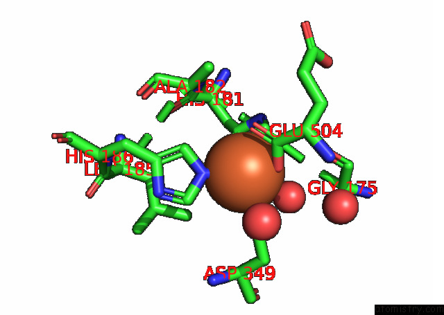
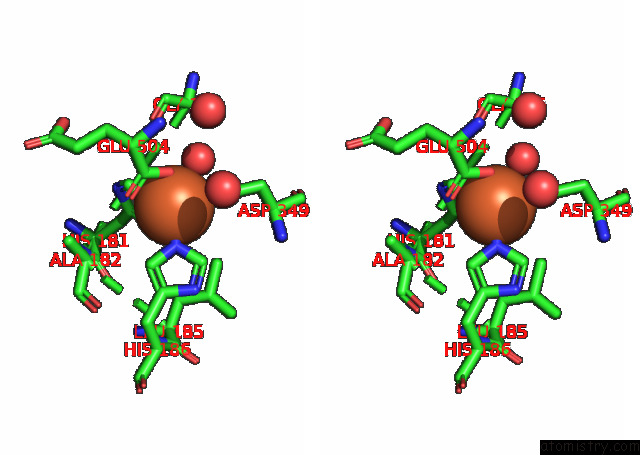
Iron binding site 1 out of 3 in 7fhr
Go back to
Iron binding site 1 out
of 3 in the Crystal Structure of A Rieske Oxygenase From Cupriavidus Metallidurans
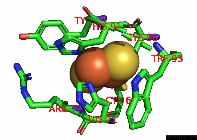
Mono view
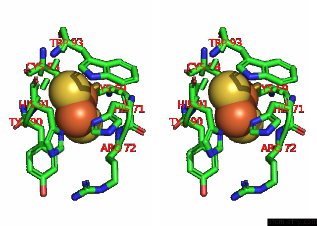
Stereo pair view
Mono view
Stereo pair view
A full contact list of Iron with other atoms in the Fe binding
site number 1 of Crystal Structure of A Rieske Oxygenase From Cupriavidus Metallidurans within 5.0Å range:
|
Iron binding site 2 out of 3 in 7fhr
Go back to
Iron binding site 2 out
of 3 in the Crystal Structure of A Rieske Oxygenase From Cupriavidus Metallidurans
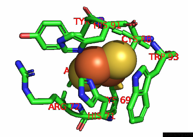
Mono view
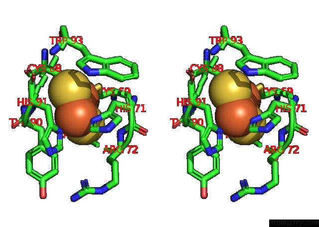
Stereo pair view
Mono view
Stereo pair view
A full contact list of Iron with other atoms in the Fe binding
site number 2 of Crystal Structure of A Rieske Oxygenase From Cupriavidus Metallidurans within 5.0Å range:
|
Iron binding site 3 out of 3 in 7fhr
Go back to
Iron binding site 3 out
of 3 in the Crystal Structure of A Rieske Oxygenase From Cupriavidus Metallidurans
Mono view
Stereo pair view
Mono view
Stereo pair view
A full contact list of Iron with other atoms in the Fe binding
site number 3 of Crystal Structure of A Rieske Oxygenase From Cupriavidus Metallidurans within 5.0Å range:
|
Reference:
J.K.Mahto,
N.Neetu,
B.Waghmode,
E.Kuatsjah,
M.Sharma,
D.Sircar,
A.K.Sharma,
S.Tomar,
L.D.Eltis,
P.Kumar.
Molecular Insights Into Substrate Recognition and Catalysis By Phthalate Dioxygenase From Comamonas Testosteroni. J.Biol.Chem. 01416 2021.
ISSN: ESSN 1083-351X
PubMed: 34800435
DOI: 10.1016/J.JBC.2021.101416
Page generated: Wed Aug 6 22:25:51 2025
ISSN: ESSN 1083-351X
PubMed: 34800435
DOI: 10.1016/J.JBC.2021.101416
Last articles
Fe in 7O3HFe in 7O4J
Fe in 7O4I
Fe in 7O3C
Fe in 7O37
Fe in 7O3E
Fe in 7O26
Fe in 7O38
Fe in 7O1V
Fe in 7O2G