Iron in PDB 7jz6: The Cryo-Em Structure of the Catalase-Peroxidase From Escherichia Coli
Enzymatic activity of The Cryo-Em Structure of the Catalase-Peroxidase From Escherichia Coli
All present enzymatic activity of The Cryo-Em Structure of the Catalase-Peroxidase From Escherichia Coli:
1.11.1.21;
Iron Binding Sites:
The binding sites of Iron atom in the The Cryo-Em Structure of the Catalase-Peroxidase From Escherichia Coli
(pdb code 7jz6). This binding sites where shown within
5.0 Angstroms radius around Iron atom.
In total 4 binding sites of Iron where determined in the
The Cryo-Em Structure of the Catalase-Peroxidase From Escherichia Coli, PDB code: 7jz6:
Jump to Iron binding site number:
1;
2;
3;
4;
Iron binding site 1 out
of 4 in 7jz6
Go back to
Iron Binding Sites List in 7jz6
Iron binding site 1 out
of 4 in the The Cryo-Em Structure of the Catalase-Peroxidase From Escherichia Coli
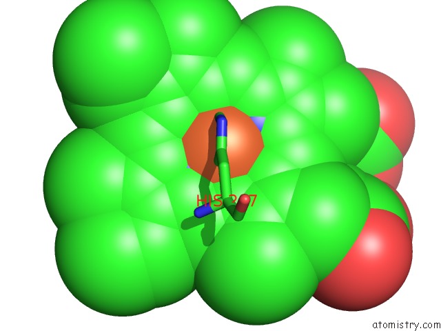
Mono view
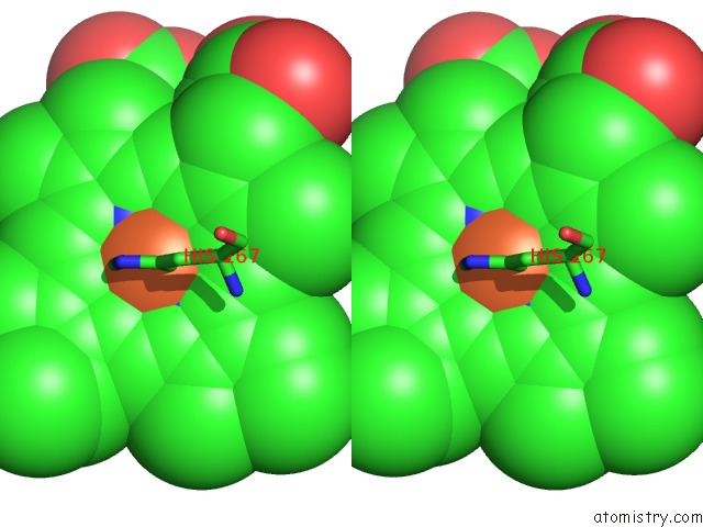
Stereo pair view
|
A full contact list of Iron with other atoms in the Fe binding
site number 1 of The Cryo-Em Structure of the Catalase-Peroxidase From Escherichia Coli within 5.0Å range:
probe
|
atom
|
residue
|
distance (Å)
|
B
|
Occ
|
A:Fe1001
b:0.0
occ:1.00
|
FE
|
A:HEM1001
|
0.0
|
0.0
|
1.0
|
NC
|
A:HEM1001
|
1.9
|
0.0
|
1.0
|
NA
|
A:HEM1001
|
2.1
|
0.0
|
1.0
|
ND
|
A:HEM1001
|
2.1
|
0.0
|
1.0
|
NB
|
A:HEM1001
|
2.1
|
0.0
|
1.0
|
NE2
|
A:HIS267
|
2.5
|
0.0
|
1.0
|
C4C
|
A:HEM1001
|
3.0
|
0.0
|
1.0
|
C1C
|
A:HEM1001
|
3.0
|
0.0
|
1.0
|
C1D
|
A:HEM1001
|
3.0
|
0.0
|
1.0
|
C4D
|
A:HEM1001
|
3.1
|
0.0
|
1.0
|
C1A
|
A:HEM1001
|
3.1
|
0.0
|
1.0
|
C4A
|
A:HEM1001
|
3.1
|
0.0
|
1.0
|
C4B
|
A:HEM1001
|
3.1
|
0.0
|
1.0
|
C1B
|
A:HEM1001
|
3.1
|
0.0
|
1.0
|
O
|
A:HOH1103
|
3.1
|
0.0
|
1.0
|
CD2
|
A:HIS267
|
3.3
|
0.0
|
1.0
|
CHD
|
A:HEM1001
|
3.4
|
0.0
|
1.0
|
CHA
|
A:HEM1001
|
3.4
|
0.0
|
1.0
|
CHC
|
A:HEM1001
|
3.4
|
0.0
|
1.0
|
CHB
|
A:HEM1001
|
3.5
|
0.0
|
1.0
|
CE1
|
A:HIS267
|
3.5
|
0.0
|
1.0
|
C3C
|
A:HEM1001
|
4.2
|
0.0
|
1.0
|
C2C
|
A:HEM1001
|
4.2
|
0.0
|
1.0
|
C2D
|
A:HEM1001
|
4.2
|
0.0
|
1.0
|
C3D
|
A:HEM1001
|
4.3
|
0.0
|
1.0
|
C3A
|
A:HEM1001
|
4.3
|
0.0
|
1.0
|
C2A
|
A:HEM1001
|
4.3
|
0.0
|
1.0
|
C3B
|
A:HEM1001
|
4.3
|
0.0
|
1.0
|
C2B
|
A:HEM1001
|
4.3
|
0.0
|
1.0
|
CG
|
A:HIS267
|
4.5
|
0.0
|
1.0
|
ND1
|
A:HIS267
|
4.6
|
0.0
|
1.0
|
NE1
|
A:TRP105
|
4.7
|
0.0
|
1.0
|
O
|
A:HOH1105
|
4.7
|
0.0
|
1.0
|
CD1
|
A:TRP105
|
4.9
|
0.0
|
1.0
|
O
|
A:HOH1102
|
5.0
|
0.0
|
1.0
|
|
Iron binding site 2 out
of 4 in 7jz6
Go back to
Iron Binding Sites List in 7jz6
Iron binding site 2 out
of 4 in the The Cryo-Em Structure of the Catalase-Peroxidase From Escherichia Coli
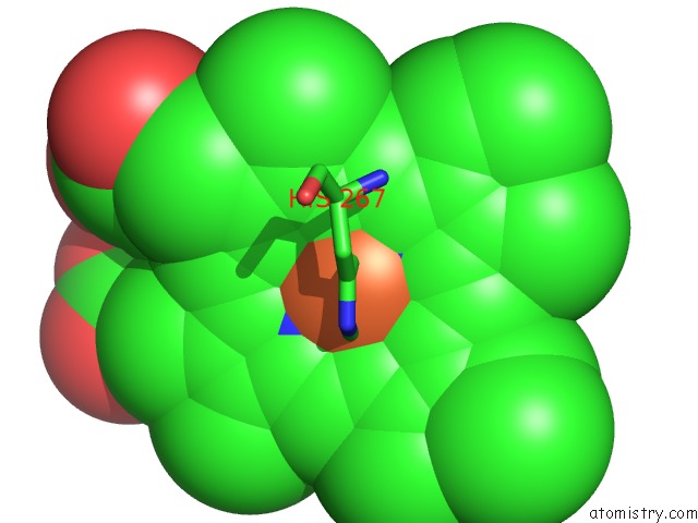
Mono view
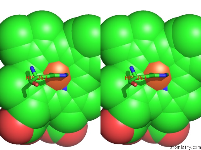
Stereo pair view
|
A full contact list of Iron with other atoms in the Fe binding
site number 2 of The Cryo-Em Structure of the Catalase-Peroxidase From Escherichia Coli within 5.0Å range:
probe
|
atom
|
residue
|
distance (Å)
|
B
|
Occ
|
B:Fe1001
b:0.0
occ:1.00
|
FE
|
B:HEM1001
|
0.0
|
0.0
|
1.0
|
NC
|
B:HEM1001
|
1.9
|
0.0
|
1.0
|
ND
|
B:HEM1001
|
2.1
|
0.0
|
1.0
|
NA
|
B:HEM1001
|
2.1
|
0.0
|
1.0
|
NB
|
B:HEM1001
|
2.1
|
0.0
|
1.0
|
NE2
|
B:HIS267
|
2.5
|
0.0
|
1.0
|
C4C
|
B:HEM1001
|
3.0
|
0.0
|
1.0
|
C1C
|
B:HEM1001
|
3.0
|
0.0
|
1.0
|
C1D
|
B:HEM1001
|
3.0
|
0.0
|
1.0
|
C4D
|
B:HEM1001
|
3.1
|
0.0
|
1.0
|
C4A
|
B:HEM1001
|
3.1
|
0.0
|
1.0
|
C4B
|
B:HEM1001
|
3.1
|
0.0
|
1.0
|
C1B
|
B:HEM1001
|
3.1
|
0.0
|
1.0
|
C1A
|
B:HEM1001
|
3.1
|
0.0
|
1.0
|
O
|
B:HOH1102
|
3.1
|
0.0
|
1.0
|
CD2
|
B:HIS267
|
3.4
|
0.0
|
1.0
|
CHD
|
B:HEM1001
|
3.4
|
0.0
|
1.0
|
CHC
|
B:HEM1001
|
3.4
|
0.0
|
1.0
|
CHA
|
B:HEM1001
|
3.4
|
0.0
|
1.0
|
CHB
|
B:HEM1001
|
3.4
|
0.0
|
1.0
|
CE1
|
B:HIS267
|
3.5
|
0.0
|
1.0
|
C3C
|
B:HEM1001
|
4.2
|
0.0
|
1.0
|
C2C
|
B:HEM1001
|
4.2
|
0.0
|
1.0
|
C2D
|
B:HEM1001
|
4.2
|
0.0
|
1.0
|
C3D
|
B:HEM1001
|
4.3
|
0.0
|
1.0
|
C3A
|
B:HEM1001
|
4.3
|
0.0
|
1.0
|
C3B
|
B:HEM1001
|
4.3
|
0.0
|
1.0
|
C2B
|
B:HEM1001
|
4.3
|
0.0
|
1.0
|
C2A
|
B:HEM1001
|
4.3
|
0.0
|
1.0
|
CG
|
B:HIS267
|
4.6
|
0.0
|
1.0
|
ND1
|
B:HIS267
|
4.6
|
0.0
|
1.0
|
NE1
|
B:TRP105
|
4.7
|
0.0
|
1.0
|
O
|
B:HOH1104
|
4.7
|
0.0
|
1.0
|
CD1
|
B:TRP105
|
4.8
|
0.0
|
1.0
|
O
|
B:HOH1101
|
5.0
|
0.0
|
1.0
|
|
Iron binding site 3 out
of 4 in 7jz6
Go back to
Iron Binding Sites List in 7jz6
Iron binding site 3 out
of 4 in the The Cryo-Em Structure of the Catalase-Peroxidase From Escherichia Coli
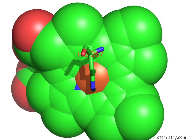
Mono view
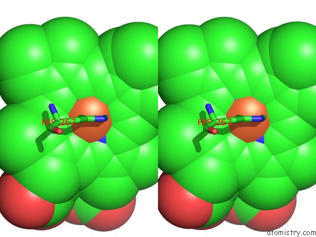
Stereo pair view
|
A full contact list of Iron with other atoms in the Fe binding
site number 3 of The Cryo-Em Structure of the Catalase-Peroxidase From Escherichia Coli within 5.0Å range:
probe
|
atom
|
residue
|
distance (Å)
|
B
|
Occ
|
C:Fe1001
b:0.0
occ:1.00
|
FE
|
C:HEM1001
|
0.0
|
0.0
|
1.0
|
NC
|
C:HEM1001
|
2.0
|
0.0
|
1.0
|
ND
|
C:HEM1001
|
2.1
|
0.0
|
1.0
|
NA
|
C:HEM1001
|
2.1
|
0.0
|
1.0
|
NB
|
C:HEM1001
|
2.1
|
0.0
|
1.0
|
NE2
|
C:HIS267
|
2.5
|
0.0
|
1.0
|
C4C
|
C:HEM1001
|
3.0
|
0.0
|
1.0
|
C1C
|
C:HEM1001
|
3.0
|
0.0
|
1.0
|
C1D
|
C:HEM1001
|
3.0
|
0.0
|
1.0
|
C4D
|
C:HEM1001
|
3.1
|
0.0
|
1.0
|
C4A
|
C:HEM1001
|
3.1
|
0.0
|
1.0
|
C4B
|
C:HEM1001
|
3.1
|
0.0
|
1.0
|
C1A
|
C:HEM1001
|
3.1
|
0.0
|
1.0
|
C1B
|
C:HEM1001
|
3.1
|
0.0
|
1.0
|
O
|
C:HOH1102
|
3.1
|
0.0
|
1.0
|
CD2
|
C:HIS267
|
3.3
|
0.0
|
1.0
|
CHD
|
C:HEM1001
|
3.4
|
0.0
|
1.0
|
CHC
|
C:HEM1001
|
3.4
|
0.0
|
1.0
|
CHB
|
C:HEM1001
|
3.4
|
0.0
|
1.0
|
CHA
|
C:HEM1001
|
3.4
|
0.0
|
1.0
|
CE1
|
C:HIS267
|
3.5
|
0.0
|
1.0
|
C3C
|
C:HEM1001
|
4.2
|
0.0
|
1.0
|
C2C
|
C:HEM1001
|
4.2
|
0.0
|
1.0
|
C2D
|
C:HEM1001
|
4.2
|
0.0
|
1.0
|
C3D
|
C:HEM1001
|
4.3
|
0.0
|
1.0
|
C3A
|
C:HEM1001
|
4.3
|
0.0
|
1.0
|
C2A
|
C:HEM1001
|
4.3
|
0.0
|
1.0
|
C3B
|
C:HEM1001
|
4.3
|
0.0
|
1.0
|
C2B
|
C:HEM1001
|
4.3
|
0.0
|
1.0
|
CG
|
C:HIS267
|
4.6
|
0.0
|
1.0
|
NE1
|
C:TRP105
|
4.6
|
0.0
|
1.0
|
ND1
|
C:HIS267
|
4.6
|
0.0
|
1.0
|
CD1
|
C:TRP105
|
4.7
|
0.0
|
1.0
|
O
|
C:HOH1104
|
4.7
|
0.0
|
1.0
|
O
|
C:HOH1101
|
5.0
|
0.0
|
1.0
|
|
Iron binding site 4 out
of 4 in 7jz6
Go back to
Iron Binding Sites List in 7jz6
Iron binding site 4 out
of 4 in the The Cryo-Em Structure of the Catalase-Peroxidase From Escherichia Coli
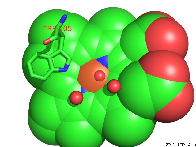
Mono view
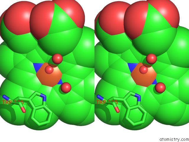
Stereo pair view
|
A full contact list of Iron with other atoms in the Fe binding
site number 4 of The Cryo-Em Structure of the Catalase-Peroxidase From Escherichia Coli within 5.0Å range:
probe
|
atom
|
residue
|
distance (Å)
|
B
|
Occ
|
D:Fe1001
b:0.0
occ:1.00
|
FE
|
D:HEM1001
|
0.0
|
0.0
|
1.0
|
NC
|
D:HEM1001
|
2.0
|
0.0
|
1.0
|
ND
|
D:HEM1001
|
2.1
|
0.0
|
1.0
|
NA
|
D:HEM1001
|
2.1
|
0.0
|
1.0
|
NB
|
D:HEM1001
|
2.1
|
0.0
|
1.0
|
NE2
|
D:HIS267
|
2.5
|
0.0
|
1.0
|
C4C
|
D:HEM1001
|
3.0
|
0.0
|
1.0
|
C1C
|
D:HEM1001
|
3.0
|
0.0
|
1.0
|
C1D
|
D:HEM1001
|
3.0
|
0.0
|
1.0
|
C4D
|
D:HEM1001
|
3.1
|
0.0
|
1.0
|
C4A
|
D:HEM1001
|
3.1
|
0.0
|
1.0
|
C1A
|
D:HEM1001
|
3.1
|
0.0
|
1.0
|
C1B
|
D:HEM1001
|
3.1
|
0.0
|
1.0
|
C4B
|
D:HEM1001
|
3.1
|
0.0
|
1.0
|
O
|
D:HOH1102
|
3.1
|
0.0
|
1.0
|
CD2
|
D:HIS267
|
3.3
|
0.0
|
1.0
|
CHD
|
D:HEM1001
|
3.4
|
0.0
|
1.0
|
CHA
|
D:HEM1001
|
3.4
|
0.0
|
1.0
|
CHC
|
D:HEM1001
|
3.4
|
0.0
|
1.0
|
CHB
|
D:HEM1001
|
3.4
|
0.0
|
1.0
|
CE1
|
D:HIS267
|
3.5
|
0.0
|
1.0
|
C3C
|
D:HEM1001
|
4.2
|
0.0
|
1.0
|
C2C
|
D:HEM1001
|
4.2
|
0.0
|
1.0
|
C2D
|
D:HEM1001
|
4.2
|
0.0
|
1.0
|
C3D
|
D:HEM1001
|
4.3
|
0.0
|
1.0
|
C3A
|
D:HEM1001
|
4.3
|
0.0
|
1.0
|
C2A
|
D:HEM1001
|
4.3
|
0.0
|
1.0
|
C2B
|
D:HEM1001
|
4.3
|
0.0
|
1.0
|
C3B
|
D:HEM1001
|
4.3
|
0.0
|
1.0
|
CG
|
D:HIS267
|
4.5
|
0.0
|
1.0
|
ND1
|
D:HIS267
|
4.6
|
0.0
|
1.0
|
NE1
|
D:TRP105
|
4.6
|
0.0
|
1.0
|
O
|
D:HOH1104
|
4.7
|
0.0
|
1.0
|
CD1
|
D:TRP105
|
4.7
|
0.0
|
1.0
|
O
|
D:HOH1101
|
5.0
|
0.0
|
1.0
|
|
Reference:
C.C.Su,
M.Lyu,
C.E.Morgan,
J.R.Bolla,
C.V.Robinson,
E.W.Yu.
A 'Build and Retrieve' Methodology to Simultaneously Solve Cryo-Em Structures of Membrane Proteins. Nat.Methods V. 18 69 2021.
ISSN: ESSN 1548-7105
PubMed: 33408407
DOI: 10.1038/S41592-020-01021-2
Page generated: Thu Aug 8 05:58:38 2024
|