Iron »
PDB 7k5g-7kvo »
7kqt »
Iron in PDB 7kqt: A 1.84-A Resolution Crystal Structure of Heme-Dependent L-Tyrosine Hydroxylase in Complex with 3-Fluoro-L-Tyrosine and Cyanide
Protein crystallography data
The structure of A 1.84-A Resolution Crystal Structure of Heme-Dependent L-Tyrosine Hydroxylase in Complex with 3-Fluoro-L-Tyrosine and Cyanide, PDB code: 7kqt
was solved by
Y.Wang,
A.Liu,
with X-Ray Crystallography technique. A brief refinement statistics is given in the table below:
| Resolution Low / High (Å) | 48.27 / 1.84 |
| Space group | P 1 21 1 |
| Cell size a, b, c (Å), α, β, γ (°) | 47.375, 129.446, 48.401, 90, 94.29, 90 |
| R / Rfree (%) | 20.3 / 25 |
Other elements in 7kqt:
The structure of A 1.84-A Resolution Crystal Structure of Heme-Dependent L-Tyrosine Hydroxylase in Complex with 3-Fluoro-L-Tyrosine and Cyanide also contains other interesting chemical elements:
| Fluorine | (F) | 4 atoms |
Iron Binding Sites:
The binding sites of Iron atom in the A 1.84-A Resolution Crystal Structure of Heme-Dependent L-Tyrosine Hydroxylase in Complex with 3-Fluoro-L-Tyrosine and Cyanide
(pdb code 7kqt). This binding sites where shown within
5.0 Angstroms radius around Iron atom.
In total 2 binding sites of Iron where determined in the A 1.84-A Resolution Crystal Structure of Heme-Dependent L-Tyrosine Hydroxylase in Complex with 3-Fluoro-L-Tyrosine and Cyanide, PDB code: 7kqt:
Jump to Iron binding site number: 1; 2;
In total 2 binding sites of Iron where determined in the A 1.84-A Resolution Crystal Structure of Heme-Dependent L-Tyrosine Hydroxylase in Complex with 3-Fluoro-L-Tyrosine and Cyanide, PDB code: 7kqt:
Jump to Iron binding site number: 1; 2;
Iron binding site 1 out of 2 in 7kqt
Go back to
Iron binding site 1 out
of 2 in the A 1.84-A Resolution Crystal Structure of Heme-Dependent L-Tyrosine Hydroxylase in Complex with 3-Fluoro-L-Tyrosine and Cyanide
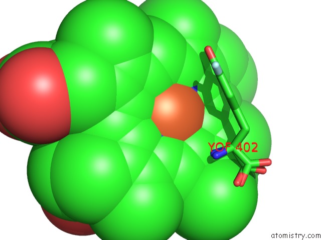
Mono view
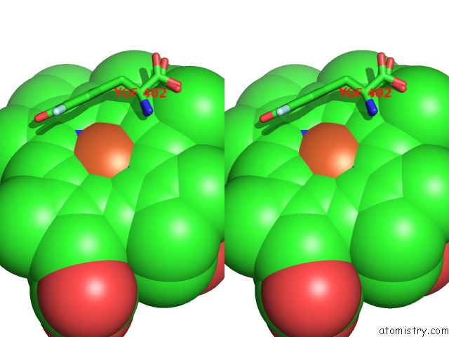
Stereo pair view
Mono view
Stereo pair view
A full contact list of Iron with other atoms in the Fe binding
site number 1 of A 1.84-A Resolution Crystal Structure of Heme-Dependent L-Tyrosine Hydroxylase in Complex with 3-Fluoro-L-Tyrosine and Cyanide within 5.0Å range:
|
Iron binding site 2 out of 2 in 7kqt
Go back to
Iron binding site 2 out
of 2 in the A 1.84-A Resolution Crystal Structure of Heme-Dependent L-Tyrosine Hydroxylase in Complex with 3-Fluoro-L-Tyrosine and Cyanide
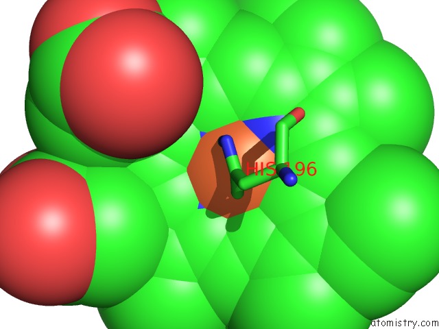
Mono view
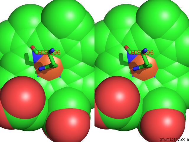
Stereo pair view
Mono view
Stereo pair view
A full contact list of Iron with other atoms in the Fe binding
site number 2 of A 1.84-A Resolution Crystal Structure of Heme-Dependent L-Tyrosine Hydroxylase in Complex with 3-Fluoro-L-Tyrosine and Cyanide within 5.0Å range:
|
Reference:
Y.Wang,
I.Davis,
I.Shin,
H.Xu,
A.Liu.
Molecular Rationale For Partitioning Between C-H and C-F Bond Activation in Heme-Dependent Tyrosine Hydroxylase. J.Am.Chem.Soc. 2021.
ISSN: ESSN 1520-5126
PubMed: 33734681
DOI: 10.1021/JACS.1C00175
Page generated: Thu Aug 8 06:38:55 2024
ISSN: ESSN 1520-5126
PubMed: 33734681
DOI: 10.1021/JACS.1C00175
Last articles
Fe in 2YXOFe in 2YRS
Fe in 2YXC
Fe in 2YNM
Fe in 2YVJ
Fe in 2YP1
Fe in 2YU2
Fe in 2YU1
Fe in 2YQB
Fe in 2YOO