Iron »
PDB 7kvp-7lvz »
7kyr »
Iron in PDB 7kyr: Crystal Structure of I107E Cub Myoglobin (I107E L29H F43H Sperm Whale Myoglobin)
Protein crystallography data
The structure of Crystal Structure of I107E Cub Myoglobin (I107E L29H F43H Sperm Whale Myoglobin), PDB code: 7kyr
was solved by
I.Petrik,
Y.Lu,
with X-Ray Crystallography technique. A brief refinement statistics is given in the table below:
| Resolution Low / High (Å) | 34.83 / 1.71 |
| Space group | P 1 21 1 |
| Cell size a, b, c (Å), α, β, γ (°) | 34.016, 31.728, 71.283, 90, 102.35, 90 |
| R / Rfree (%) | 16.6 / 21 |
Other elements in 7kyr:
The structure of Crystal Structure of I107E Cub Myoglobin (I107E L29H F43H Sperm Whale Myoglobin) also contains other interesting chemical elements:
| Sodium | (Na) | 1 atom |
Iron Binding Sites:
The binding sites of Iron atom in the Crystal Structure of I107E Cub Myoglobin (I107E L29H F43H Sperm Whale Myoglobin)
(pdb code 7kyr). This binding sites where shown within
5.0 Angstroms radius around Iron atom.
In total only one binding site of Iron was determined in the Crystal Structure of I107E Cub Myoglobin (I107E L29H F43H Sperm Whale Myoglobin), PDB code: 7kyr:
In total only one binding site of Iron was determined in the Crystal Structure of I107E Cub Myoglobin (I107E L29H F43H Sperm Whale Myoglobin), PDB code: 7kyr:
Iron binding site 1 out of 1 in 7kyr
Go back to
Iron binding site 1 out
of 1 in the Crystal Structure of I107E Cub Myoglobin (I107E L29H F43H Sperm Whale Myoglobin)
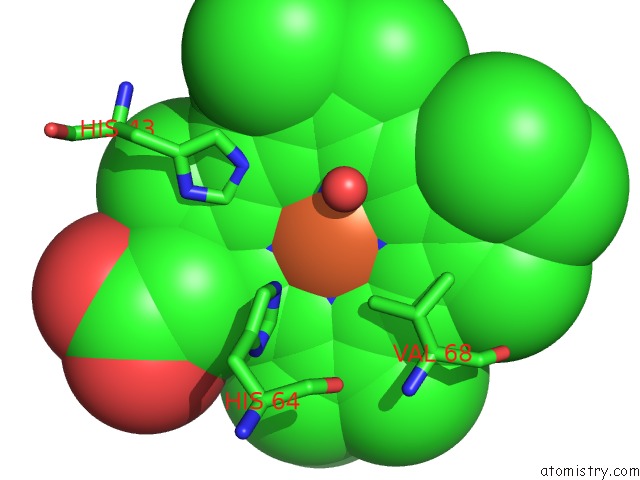
Mono view
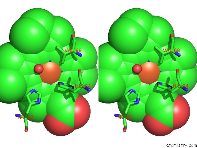
Stereo pair view
Mono view
Stereo pair view
A full contact list of Iron with other atoms in the Fe binding
site number 1 of Crystal Structure of I107E Cub Myoglobin (I107E L29H F43H Sperm Whale Myoglobin) within 5.0Å range:
|
Reference:
I.D.Petrik,
R.Davydov,
M.Kahle,
B.Sandoval,
S.Dwaraknath,
P.Adelroth,
B.Hoffman,
Y.Lu.
An Engineered Glutamate in Biosynthetic Models of Heme-Copper Oxidases Drives Complete Product Selectivity By Tuning the Hydrogen-Bonding Network. Biochemistry 2021.
ISSN: ISSN 0006-2960
PubMed: 33464878
DOI: 10.1021/ACS.BIOCHEM.0C00852
Page generated: Thu Aug 8 06:55:37 2024
ISSN: ISSN 0006-2960
PubMed: 33464878
DOI: 10.1021/ACS.BIOCHEM.0C00852
Last articles
Zn in 9J0NZn in 9J0O
Zn in 9J0P
Zn in 9FJX
Zn in 9EKB
Zn in 9C0F
Zn in 9CAH
Zn in 9CH0
Zn in 9CH3
Zn in 9CH1