Iron in PDB 7lhr: Crystal Structure of Adenosine-5'-Phosphosulfate Reductase From Mycobacterium Tuberculosis
Enzymatic activity of Crystal Structure of Adenosine-5'-Phosphosulfate Reductase From Mycobacterium Tuberculosis
All present enzymatic activity of Crystal Structure of Adenosine-5'-Phosphosulfate Reductase From Mycobacterium Tuberculosis:
1.8.4.8;
Protein crystallography data
The structure of Crystal Structure of Adenosine-5'-Phosphosulfate Reductase From Mycobacterium Tuberculosis, PDB code: 7lhr
was solved by
P.R.Feliciano,
C.L.Drennan,
with X-Ray Crystallography technique. A brief refinement statistics is given in the table below:
Resolution Low / High (Å)
|
48.25 /
3.11
|
Space group
|
P 43 21 2
|
Cell size a, b, c (Å), α, β, γ (°)
|
77.414,
77.414,
204.352,
90,
90,
90
|
R / Rfree (%)
|
20.7 /
24.9
|
Iron Binding Sites:
The binding sites of Iron atom in the Crystal Structure of Adenosine-5'-Phosphosulfate Reductase From Mycobacterium Tuberculosis
(pdb code 7lhr). This binding sites where shown within
5.0 Angstroms radius around Iron atom.
In total 8 binding sites of Iron where determined in the
Crystal Structure of Adenosine-5'-Phosphosulfate Reductase From Mycobacterium Tuberculosis, PDB code: 7lhr:
Jump to Iron binding site number:
1;
2;
3;
4;
5;
6;
7;
8;
Iron binding site 1 out
of 8 in 7lhr
Go back to
Iron Binding Sites List in 7lhr
Iron binding site 1 out
of 8 in the Crystal Structure of Adenosine-5'-Phosphosulfate Reductase From Mycobacterium Tuberculosis
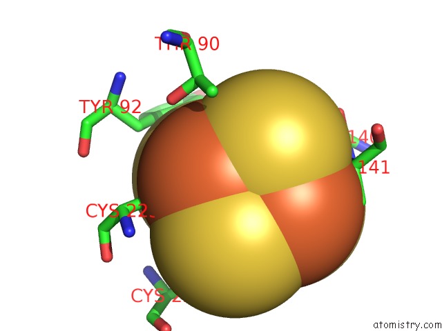
Mono view
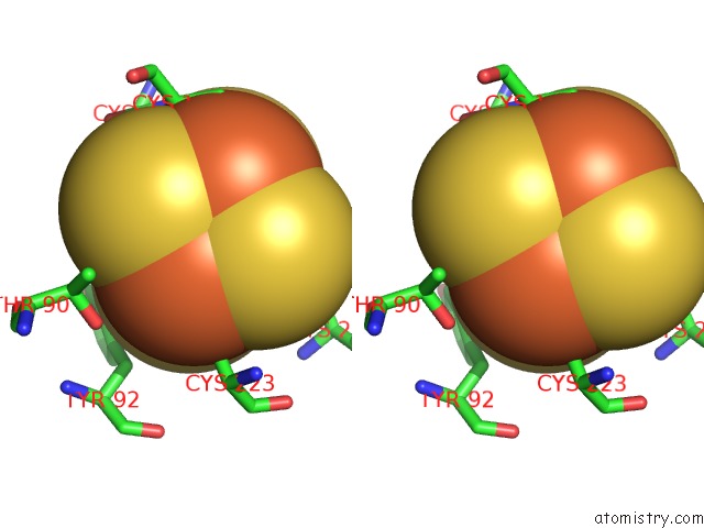
Stereo pair view
|
A full contact list of Iron with other atoms in the Fe binding
site number 1 of Crystal Structure of Adenosine-5'-Phosphosulfate Reductase From Mycobacterium Tuberculosis within 5.0Å range:
probe
|
atom
|
residue
|
distance (Å)
|
B
|
Occ
|
A:Fe301
b:87.2
occ:1.00
|
FE1
|
A:SF4301
|
0.0
|
87.2
|
1.0
|
S4
|
A:SF4301
|
2.3
|
82.2
|
1.0
|
SG
|
A:CYS223
|
2.3
|
116.0
|
1.0
|
S2
|
A:SF4301
|
2.3
|
94.3
|
1.0
|
S3
|
A:SF4301
|
2.3
|
101.2
|
1.0
|
FE2
|
A:SF4301
|
2.7
|
93.1
|
1.0
|
FE3
|
A:SF4301
|
2.7
|
87.2
|
1.0
|
FE4
|
A:SF4301
|
2.7
|
98.5
|
1.0
|
CB
|
A:CYS223
|
3.0
|
102.6
|
1.0
|
S1
|
A:SF4301
|
3.9
|
88.5
|
1.0
|
OG1
|
A:THR90
|
4.2
|
83.9
|
1.0
|
CA
|
A:CYS223
|
4.4
|
95.2
|
1.0
|
CD2
|
A:TYR92
|
4.7
|
110.8
|
1.0
|
CB
|
A:THR90
|
4.7
|
83.1
|
1.0
|
SG
|
A:CYS226
|
4.7
|
114.4
|
1.0
|
CB
|
A:TYR92
|
4.7
|
102.7
|
1.0
|
N
|
A:CYS223
|
4.8
|
100.0
|
1.0
|
CG2
|
A:THR90
|
4.8
|
80.7
|
1.0
|
SG
|
A:CYS141
|
4.8
|
99.0
|
1.0
|
SG
|
A:CYS140
|
4.9
|
98.3
|
1.0
|
|
Iron binding site 2 out
of 8 in 7lhr
Go back to
Iron Binding Sites List in 7lhr
Iron binding site 2 out
of 8 in the Crystal Structure of Adenosine-5'-Phosphosulfate Reductase From Mycobacterium Tuberculosis
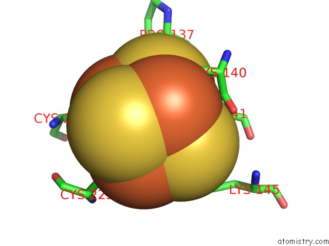
Mono view
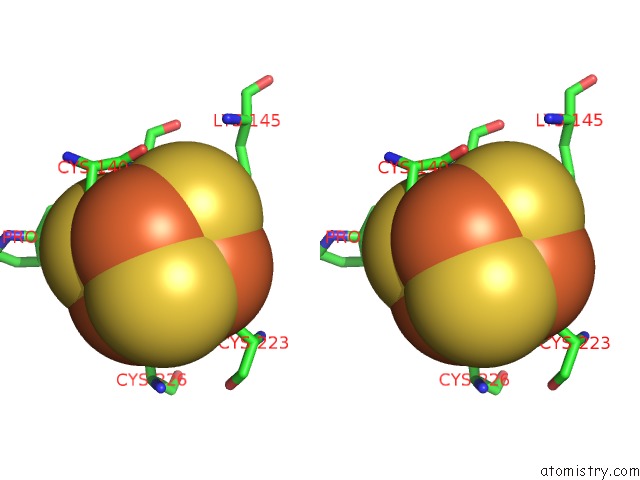
Stereo pair view
|
A full contact list of Iron with other atoms in the Fe binding
site number 2 of Crystal Structure of Adenosine-5'-Phosphosulfate Reductase From Mycobacterium Tuberculosis within 5.0Å range:
probe
|
atom
|
residue
|
distance (Å)
|
B
|
Occ
|
A:Fe301
b:93.1
occ:1.00
|
FE2
|
A:SF4301
|
0.0
|
93.1
|
1.0
|
S3
|
A:SF4301
|
2.3
|
101.2
|
1.0
|
SG
|
A:CYS141
|
2.3
|
99.0
|
1.0
|
S1
|
A:SF4301
|
2.3
|
88.5
|
1.0
|
S4
|
A:SF4301
|
2.3
|
82.2
|
1.0
|
FE4
|
A:SF4301
|
2.7
|
98.5
|
1.0
|
FE1
|
A:SF4301
|
2.7
|
87.2
|
1.0
|
FE3
|
A:SF4301
|
2.7
|
87.2
|
1.0
|
CB
|
A:CYS141
|
3.4
|
91.2
|
1.0
|
CA
|
A:CYS141
|
3.6
|
87.9
|
1.0
|
N
|
A:CYS141
|
3.8
|
80.7
|
1.0
|
S2
|
A:SF4301
|
3.9
|
94.3
|
1.0
|
CE
|
A:LYS145
|
4.3
|
109.0
|
1.0
|
O
|
A:PRO137
|
4.4
|
146.2
|
1.0
|
C
|
A:CYS140
|
4.4
|
81.0
|
1.0
|
NZ
|
A:LYS145
|
4.6
|
95.9
|
1.0
|
SG
|
A:CYS140
|
4.7
|
98.3
|
1.0
|
SG
|
A:CYS223
|
4.7
|
116.0
|
1.0
|
SG
|
A:CYS226
|
4.7
|
114.4
|
1.0
|
CB
|
A:CYS140
|
4.8
|
94.5
|
1.0
|
CD
|
A:LYS145
|
4.8
|
104.2
|
1.0
|
O
|
A:CYS140
|
4.8
|
79.5
|
1.0
|
C
|
A:CYS141
|
5.0
|
104.1
|
1.0
|
|
Iron binding site 3 out
of 8 in 7lhr
Go back to
Iron Binding Sites List in 7lhr
Iron binding site 3 out
of 8 in the Crystal Structure of Adenosine-5'-Phosphosulfate Reductase From Mycobacterium Tuberculosis
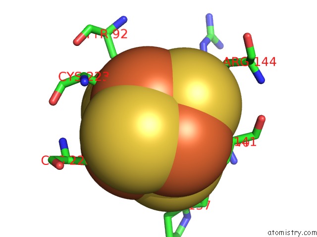
Mono view
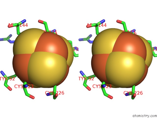
Stereo pair view
|
A full contact list of Iron with other atoms in the Fe binding
site number 3 of Crystal Structure of Adenosine-5'-Phosphosulfate Reductase From Mycobacterium Tuberculosis within 5.0Å range:
probe
|
atom
|
residue
|
distance (Å)
|
B
|
Occ
|
A:Fe301
b:87.2
occ:1.00
|
FE3
|
A:SF4301
|
0.0
|
87.2
|
1.0
|
S2
|
A:SF4301
|
2.3
|
94.3
|
1.0
|
S4
|
A:SF4301
|
2.3
|
82.2
|
1.0
|
SG
|
A:CYS140
|
2.3
|
98.3
|
1.0
|
S1
|
A:SF4301
|
2.3
|
88.5
|
1.0
|
FE1
|
A:SF4301
|
2.7
|
87.2
|
1.0
|
FE2
|
A:SF4301
|
2.7
|
93.1
|
1.0
|
FE4
|
A:SF4301
|
2.7
|
98.5
|
1.0
|
CB
|
A:CYS140
|
3.2
|
94.5
|
1.0
|
S3
|
A:SF4301
|
3.9
|
101.2
|
1.0
|
CB
|
A:PHE133
|
3.9
|
124.7
|
1.0
|
C
|
A:CYS140
|
3.9
|
81.0
|
1.0
|
N
|
A:CYS141
|
4.1
|
80.7
|
1.0
|
CA
|
A:CYS140
|
4.2
|
84.7
|
1.0
|
O
|
A:CYS140
|
4.2
|
79.5
|
1.0
|
CD2
|
A:TYR92
|
4.4
|
110.8
|
1.0
|
CE2
|
A:TYR92
|
4.6
|
117.5
|
1.0
|
CA
|
A:CYS141
|
4.6
|
87.9
|
1.0
|
CA
|
A:PHE133
|
4.6
|
129.8
|
1.0
|
SG
|
A:CYS141
|
4.7
|
99.0
|
1.0
|
CD
|
A:ARG144
|
4.8
|
98.5
|
1.0
|
SG
|
A:CYS226
|
4.8
|
114.4
|
1.0
|
SG
|
A:CYS223
|
4.9
|
116.0
|
1.0
|
O
|
A:PRO137
|
4.9
|
146.2
|
1.0
|
|
Iron binding site 4 out
of 8 in 7lhr
Go back to
Iron Binding Sites List in 7lhr
Iron binding site 4 out
of 8 in the Crystal Structure of Adenosine-5'-Phosphosulfate Reductase From Mycobacterium Tuberculosis
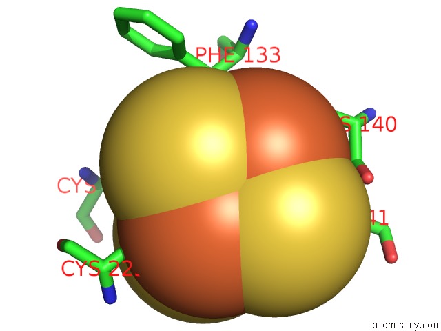
Mono view
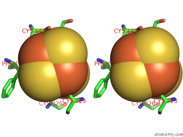
Stereo pair view
|
A full contact list of Iron with other atoms in the Fe binding
site number 4 of Crystal Structure of Adenosine-5'-Phosphosulfate Reductase From Mycobacterium Tuberculosis within 5.0Å range:
probe
|
atom
|
residue
|
distance (Å)
|
B
|
Occ
|
A:Fe301
b:98.5
occ:1.00
|
FE4
|
A:SF4301
|
0.0
|
98.5
|
1.0
|
S1
|
A:SF4301
|
2.3
|
88.5
|
1.0
|
SG
|
A:CYS226
|
2.3
|
114.4
|
1.0
|
S3
|
A:SF4301
|
2.3
|
101.2
|
1.0
|
S2
|
A:SF4301
|
2.3
|
94.3
|
1.0
|
FE2
|
A:SF4301
|
2.7
|
93.1
|
1.0
|
FE3
|
A:SF4301
|
2.7
|
87.2
|
1.0
|
FE1
|
A:SF4301
|
2.7
|
87.2
|
1.0
|
CB
|
A:CYS226
|
3.3
|
109.8
|
1.0
|
S4
|
A:SF4301
|
3.9
|
82.2
|
1.0
|
CB
|
A:PHE133
|
4.0
|
124.7
|
1.0
|
CB
|
A:CYS223
|
4.4
|
102.6
|
1.0
|
CA
|
A:CYS226
|
4.6
|
113.9
|
1.0
|
SG
|
A:CYS223
|
4.7
|
116.0
|
1.0
|
SG
|
A:CYS141
|
4.7
|
99.0
|
1.0
|
N
|
A:CYS226
|
4.7
|
111.3
|
1.0
|
SG
|
A:CYS140
|
4.7
|
98.3
|
1.0
|
CG
|
A:PHE133
|
4.9
|
127.4
|
1.0
|
CA
|
A:PHE133
|
5.0
|
129.8
|
1.0
|
|
Iron binding site 5 out
of 8 in 7lhr
Go back to
Iron Binding Sites List in 7lhr
Iron binding site 5 out
of 8 in the Crystal Structure of Adenosine-5'-Phosphosulfate Reductase From Mycobacterium Tuberculosis
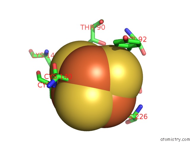
Mono view
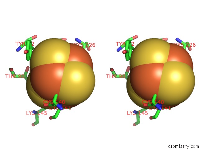
Stereo pair view
|
A full contact list of Iron with other atoms in the Fe binding
site number 5 of Crystal Structure of Adenosine-5'-Phosphosulfate Reductase From Mycobacterium Tuberculosis within 5.0Å range:
probe
|
atom
|
residue
|
distance (Å)
|
B
|
Occ
|
B:Fe301
b:86.7
occ:1.00
|
FE1
|
B:SF4301
|
0.0
|
86.7
|
1.0
|
S4
|
B:SF4301
|
2.3
|
78.4
|
1.0
|
S3
|
B:SF4301
|
2.3
|
76.3
|
1.0
|
S2
|
B:SF4301
|
2.3
|
77.1
|
1.0
|
SG
|
B:CYS223
|
2.3
|
89.5
|
1.0
|
FE2
|
B:SF4301
|
2.7
|
85.0
|
1.0
|
FE3
|
B:SF4301
|
2.7
|
81.5
|
1.0
|
FE4
|
B:SF4301
|
2.7
|
74.6
|
1.0
|
CB
|
B:CYS223
|
3.0
|
77.4
|
1.0
|
S1
|
B:SF4301
|
3.9
|
83.6
|
1.0
|
CE
|
B:LYS145
|
4.0
|
117.8
|
1.0
|
OG1
|
B:THR90
|
4.2
|
67.5
|
1.0
|
NZ
|
B:LYS145
|
4.3
|
121.7
|
1.0
|
CA
|
B:CYS223
|
4.4
|
65.3
|
1.0
|
CB
|
B:THR90
|
4.6
|
68.5
|
1.0
|
SG
|
B:CYS226
|
4.7
|
72.9
|
1.0
|
CG2
|
B:THR90
|
4.7
|
68.3
|
1.0
|
CB
|
B:TYR92
|
4.8
|
95.6
|
1.0
|
SG
|
B:CYS141
|
4.8
|
92.7
|
1.0
|
SG
|
B:CYS140
|
4.8
|
111.9
|
1.0
|
N
|
B:CYS223
|
4.9
|
69.0
|
1.0
|
CD2
|
B:TYR92
|
4.9
|
91.2
|
1.0
|
CD
|
B:LYS145
|
4.9
|
100.7
|
1.0
|
|
Iron binding site 6 out
of 8 in 7lhr
Go back to
Iron Binding Sites List in 7lhr
Iron binding site 6 out
of 8 in the Crystal Structure of Adenosine-5'-Phosphosulfate Reductase From Mycobacterium Tuberculosis
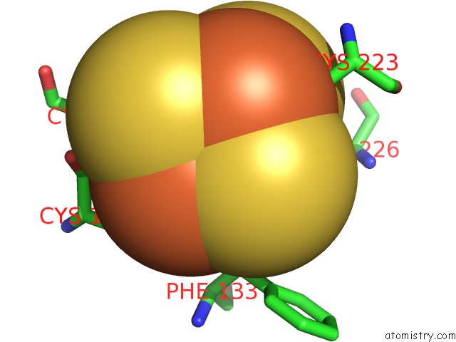
Mono view
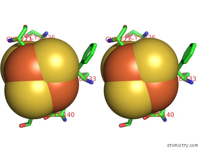
Stereo pair view
|
A full contact list of Iron with other atoms in the Fe binding
site number 6 of Crystal Structure of Adenosine-5'-Phosphosulfate Reductase From Mycobacterium Tuberculosis within 5.0Å range:
probe
|
atom
|
residue
|
distance (Å)
|
B
|
Occ
|
B:Fe301
b:85.0
occ:1.00
|
FE2
|
B:SF4301
|
0.0
|
85.0
|
1.0
|
SG
|
B:CYS226
|
2.2
|
72.9
|
1.0
|
S4
|
B:SF4301
|
2.3
|
78.4
|
1.0
|
S3
|
B:SF4301
|
2.3
|
76.3
|
1.0
|
S1
|
B:SF4301
|
2.3
|
83.6
|
1.0
|
FE1
|
B:SF4301
|
2.7
|
86.7
|
1.0
|
FE4
|
B:SF4301
|
2.7
|
74.6
|
1.0
|
FE3
|
B:SF4301
|
2.7
|
81.5
|
1.0
|
CB
|
B:CYS226
|
3.2
|
75.2
|
1.0
|
S2
|
B:SF4301
|
3.9
|
77.1
|
1.0
|
CB
|
B:PHE133
|
4.0
|
106.2
|
1.0
|
CB
|
B:CYS223
|
4.3
|
77.4
|
1.0
|
CA
|
B:CYS226
|
4.5
|
75.3
|
1.0
|
N
|
B:CYS226
|
4.6
|
72.9
|
1.0
|
SG
|
B:CYS223
|
4.6
|
89.5
|
1.0
|
SG
|
B:CYS141
|
4.7
|
92.7
|
1.0
|
SG
|
B:CYS140
|
4.7
|
111.9
|
1.0
|
CG
|
B:PHE133
|
4.9
|
106.3
|
1.0
|
|
Iron binding site 7 out
of 8 in 7lhr
Go back to
Iron Binding Sites List in 7lhr
Iron binding site 7 out
of 8 in the Crystal Structure of Adenosine-5'-Phosphosulfate Reductase From Mycobacterium Tuberculosis
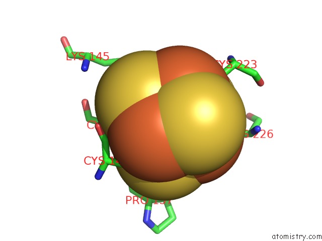
Mono view
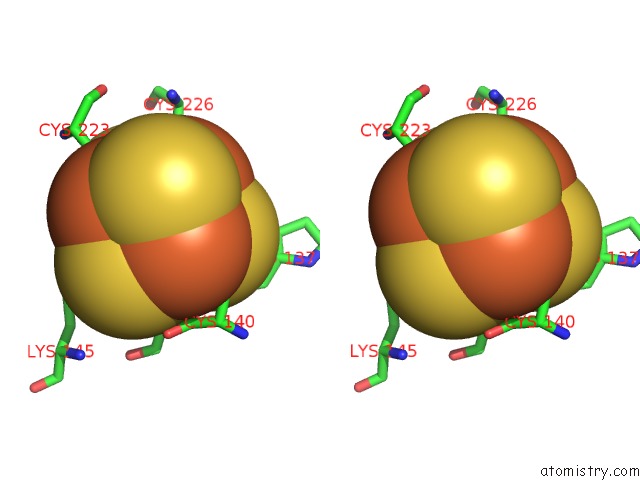
Stereo pair view
|
A full contact list of Iron with other atoms in the Fe binding
site number 7 of Crystal Structure of Adenosine-5'-Phosphosulfate Reductase From Mycobacterium Tuberculosis within 5.0Å range:
probe
|
atom
|
residue
|
distance (Å)
|
B
|
Occ
|
B:Fe301
b:81.5
occ:1.00
|
FE3
|
B:SF4301
|
0.0
|
81.5
|
1.0
|
SG
|
B:CYS141
|
2.3
|
92.7
|
1.0
|
S2
|
B:SF4301
|
2.3
|
77.1
|
1.0
|
S1
|
B:SF4301
|
2.3
|
83.6
|
1.0
|
S4
|
B:SF4301
|
2.3
|
78.4
|
1.0
|
FE4
|
B:SF4301
|
2.7
|
74.6
|
1.0
|
FE1
|
B:SF4301
|
2.7
|
86.7
|
1.0
|
FE2
|
B:SF4301
|
2.7
|
85.0
|
1.0
|
CB
|
B:CYS141
|
3.5
|
90.0
|
1.0
|
CA
|
B:CYS141
|
3.6
|
82.7
|
1.0
|
N
|
B:CYS141
|
3.8
|
86.3
|
1.0
|
S3
|
B:SF4301
|
3.9
|
76.3
|
1.0
|
CE
|
B:LYS145
|
4.4
|
117.8
|
1.0
|
C
|
B:CYS140
|
4.4
|
96.6
|
1.0
|
O
|
B:PRO137
|
4.5
|
110.0
|
1.0
|
NZ
|
B:LYS145
|
4.6
|
121.7
|
1.0
|
CD
|
B:LYS145
|
4.6
|
100.7
|
1.0
|
SG
|
B:CYS140
|
4.6
|
111.9
|
1.0
|
SG
|
B:CYS226
|
4.7
|
72.9
|
1.0
|
SG
|
B:CYS223
|
4.7
|
89.5
|
1.0
|
CB
|
B:CYS140
|
4.8
|
112.3
|
1.0
|
O
|
B:CYS140
|
4.8
|
100.4
|
1.0
|
CG
|
B:LYS145
|
4.9
|
93.5
|
1.0
|
|
Iron binding site 8 out
of 8 in 7lhr
Go back to
Iron Binding Sites List in 7lhr
Iron binding site 8 out
of 8 in the Crystal Structure of Adenosine-5'-Phosphosulfate Reductase From Mycobacterium Tuberculosis
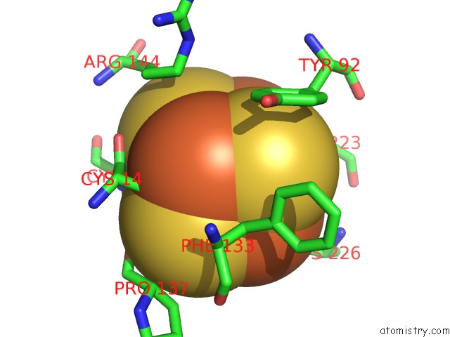
Mono view
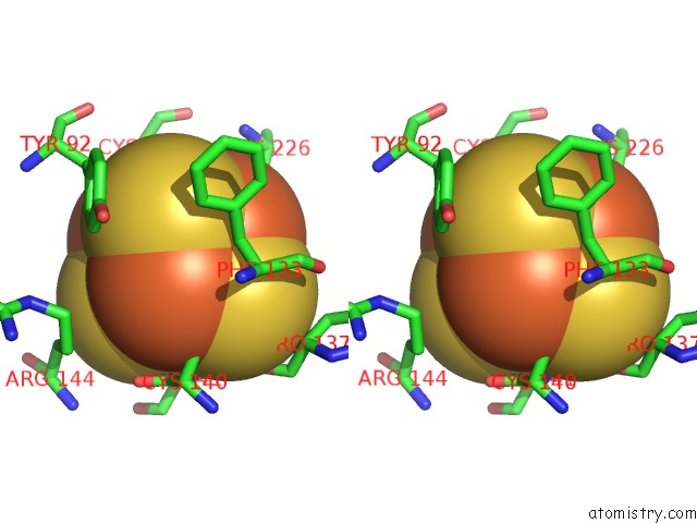
Stereo pair view
|
A full contact list of Iron with other atoms in the Fe binding
site number 8 of Crystal Structure of Adenosine-5'-Phosphosulfate Reductase From Mycobacterium Tuberculosis within 5.0Å range:
probe
|
atom
|
residue
|
distance (Å)
|
B
|
Occ
|
B:Fe301
b:74.6
occ:1.00
|
FE4
|
B:SF4301
|
0.0
|
74.6
|
1.0
|
SG
|
B:CYS140
|
2.3
|
111.9
|
1.0
|
S1
|
B:SF4301
|
2.3
|
83.6
|
1.0
|
S2
|
B:SF4301
|
2.3
|
77.1
|
1.0
|
S3
|
B:SF4301
|
2.3
|
76.3
|
1.0
|
FE3
|
B:SF4301
|
2.7
|
81.5
|
1.0
|
FE2
|
B:SF4301
|
2.7
|
85.0
|
1.0
|
FE1
|
B:SF4301
|
2.7
|
86.7
|
1.0
|
CB
|
B:CYS140
|
3.2
|
112.3
|
1.0
|
S4
|
B:SF4301
|
3.9
|
78.4
|
1.0
|
C
|
B:CYS140
|
3.9
|
96.6
|
1.0
|
CB
|
B:PHE133
|
4.0
|
106.2
|
1.0
|
N
|
B:CYS141
|
4.1
|
86.3
|
1.0
|
CA
|
B:CYS140
|
4.2
|
106.4
|
1.0
|
O
|
B:CYS140
|
4.2
|
100.4
|
1.0
|
CA
|
B:CYS141
|
4.6
|
82.7
|
1.0
|
CD2
|
B:TYR92
|
4.6
|
91.2
|
1.0
|
SG
|
B:CYS141
|
4.7
|
92.7
|
1.0
|
CA
|
B:PHE133
|
4.7
|
105.8
|
1.0
|
CD
|
B:ARG144
|
4.7
|
77.8
|
1.0
|
SG
|
B:CYS226
|
4.8
|
72.9
|
1.0
|
SG
|
B:CYS223
|
4.9
|
89.5
|
1.0
|
CE2
|
B:TYR92
|
4.9
|
94.6
|
1.0
|
O
|
B:PRO137
|
4.9
|
110.0
|
1.0
|
|
Reference:
P.R.Feliciano,
K.S.Carroll,
C.L.Drennan.
Crystal Structure of the [4FE-4S] Cluster-Containing Adenosine-5'-Phosphosulfate Reductase From Mycobacterium Tuberculosis . Acs Omega V. 6 13756 2021.
ISSN: ESSN 2470-1343
PubMed: 34095667
DOI: 10.1021/ACSOMEGA.1C01043
Page generated: Thu Aug 8 06:58:31 2024
|