Iron »
PDB 7kvp-7lvz »
7lhs »
Iron in PDB 7lhs: Crystal Structure of Adenosine-5'-Phosphosulfate Reductase From Mycobacterium Tuberculosis in A Complex with Substrate Aps
Enzymatic activity of Crystal Structure of Adenosine-5'-Phosphosulfate Reductase From Mycobacterium Tuberculosis in A Complex with Substrate Aps
All present enzymatic activity of Crystal Structure of Adenosine-5'-Phosphosulfate Reductase From Mycobacterium Tuberculosis in A Complex with Substrate Aps:
1.8.4.8;
1.8.4.8;
Protein crystallography data
The structure of Crystal Structure of Adenosine-5'-Phosphosulfate Reductase From Mycobacterium Tuberculosis in A Complex with Substrate Aps, PDB code: 7lhs
was solved by
P.R.Feliciano,
C.L.Drennan,
with X-Ray Crystallography technique. A brief refinement statistics is given in the table below:
| Resolution Low / High (Å) | 48.34 / 3.11 |
| Space group | P 43 21 2 |
| Cell size a, b, c (Å), α, β, γ (°) | 77.502, 77.502, 205.31, 90, 90, 90 |
| R / Rfree (%) | 21.9 / 26.4 |
Iron Binding Sites:
The binding sites of Iron atom in the Crystal Structure of Adenosine-5'-Phosphosulfate Reductase From Mycobacterium Tuberculosis in A Complex with Substrate Aps
(pdb code 7lhs). This binding sites where shown within
5.0 Angstroms radius around Iron atom.
In total 8 binding sites of Iron where determined in the Crystal Structure of Adenosine-5'-Phosphosulfate Reductase From Mycobacterium Tuberculosis in A Complex with Substrate Aps, PDB code: 7lhs:
Jump to Iron binding site number: 1; 2; 3; 4; 5; 6; 7; 8;
In total 8 binding sites of Iron where determined in the Crystal Structure of Adenosine-5'-Phosphosulfate Reductase From Mycobacterium Tuberculosis in A Complex with Substrate Aps, PDB code: 7lhs:
Jump to Iron binding site number: 1; 2; 3; 4; 5; 6; 7; 8;
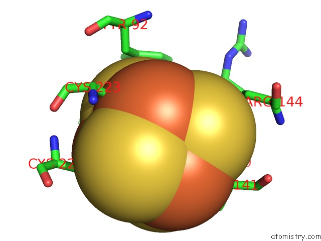
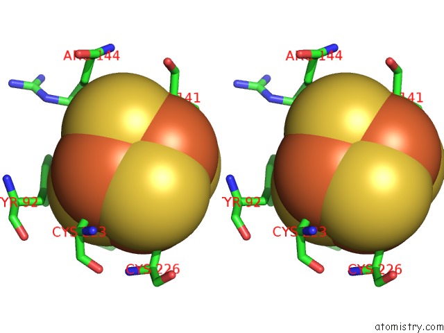
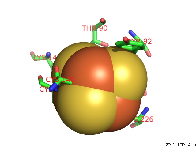
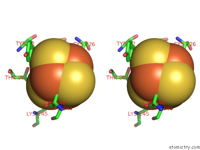
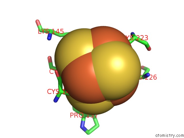
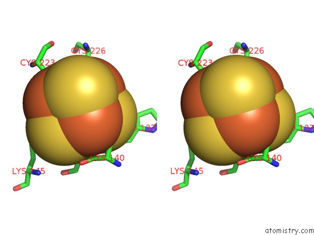
Iron binding site 1 out of 8 in 7lhs
Go back to
Iron binding site 1 out
of 8 in the Crystal Structure of Adenosine-5'-Phosphosulfate Reductase From Mycobacterium Tuberculosis in A Complex with Substrate Aps
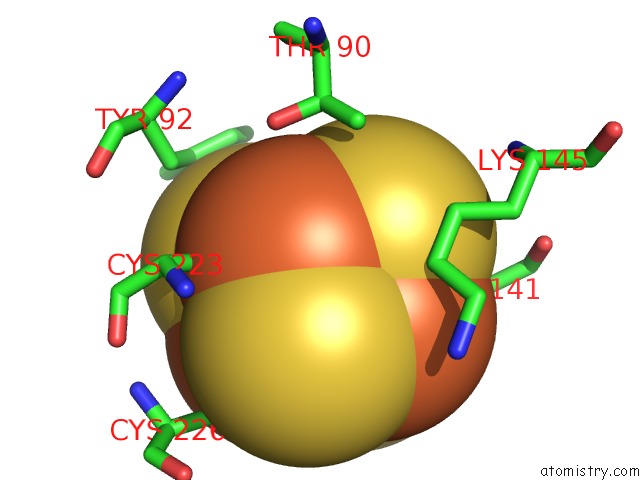
Mono view
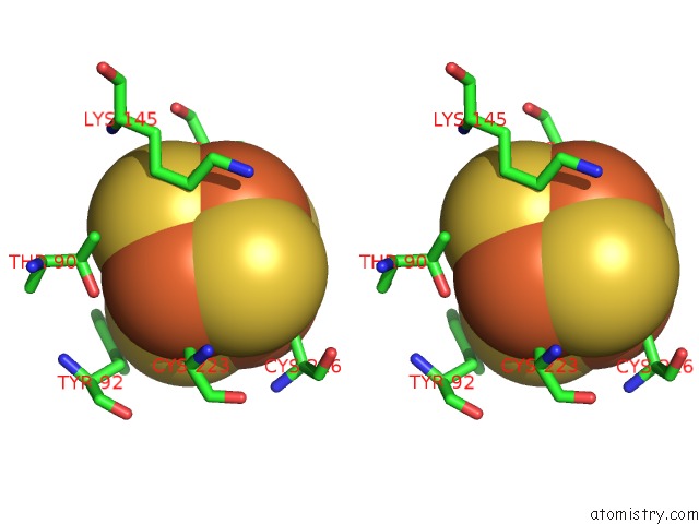
Stereo pair view
Mono view
Stereo pair view
A full contact list of Iron with other atoms in the Fe binding
site number 1 of Crystal Structure of Adenosine-5'-Phosphosulfate Reductase From Mycobacterium Tuberculosis in A Complex with Substrate Aps within 5.0Å range:
|
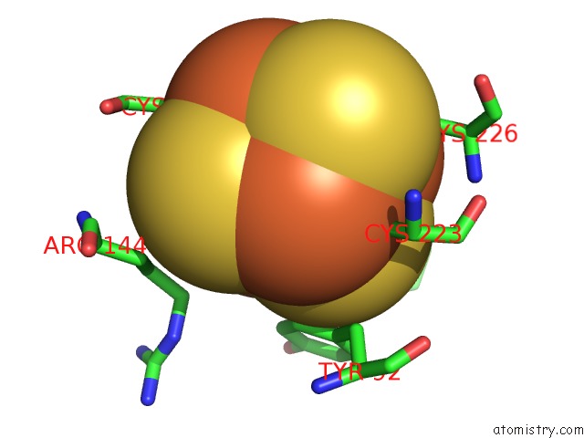
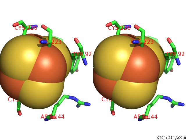
Iron binding site 2 out of 8 in 7lhs
Go back to
Iron binding site 2 out
of 8 in the Crystal Structure of Adenosine-5'-Phosphosulfate Reductase From Mycobacterium Tuberculosis in A Complex with Substrate Aps
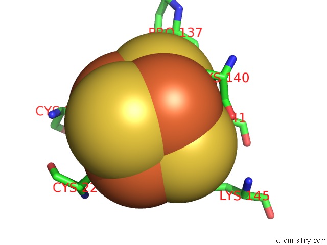
Mono view
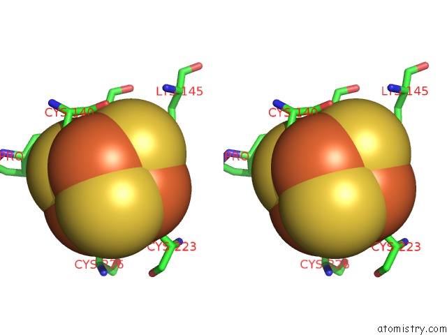
Stereo pair view
Mono view
Stereo pair view
A full contact list of Iron with other atoms in the Fe binding
site number 2 of Crystal Structure of Adenosine-5'-Phosphosulfate Reductase From Mycobacterium Tuberculosis in A Complex with Substrate Aps within 5.0Å range:
|
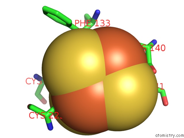
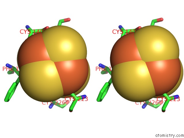
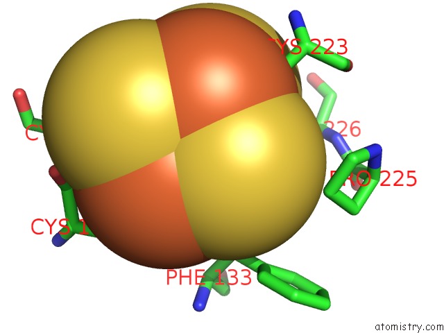
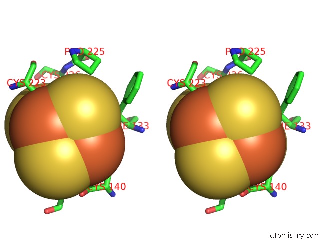
Iron binding site 3 out of 8 in 7lhs
Go back to
Iron binding site 3 out
of 8 in the Crystal Structure of Adenosine-5'-Phosphosulfate Reductase From Mycobacterium Tuberculosis in A Complex with Substrate Aps
Mono view
Stereo pair view
Mono view
Stereo pair view
A full contact list of Iron with other atoms in the Fe binding
site number 3 of Crystal Structure of Adenosine-5'-Phosphosulfate Reductase From Mycobacterium Tuberculosis in A Complex with Substrate Aps within 5.0Å range:
|
Iron binding site 4 out of 8 in 7lhs
Go back to
Iron binding site 4 out
of 8 in the Crystal Structure of Adenosine-5'-Phosphosulfate Reductase From Mycobacterium Tuberculosis in A Complex with Substrate Aps
Mono view
Stereo pair view
Mono view
Stereo pair view
A full contact list of Iron with other atoms in the Fe binding
site number 4 of Crystal Structure of Adenosine-5'-Phosphosulfate Reductase From Mycobacterium Tuberculosis in A Complex with Substrate Aps within 5.0Å range:
|
Iron binding site 5 out of 8 in 7lhs
Go back to
Iron binding site 5 out
of 8 in the Crystal Structure of Adenosine-5'-Phosphosulfate Reductase From Mycobacterium Tuberculosis in A Complex with Substrate Aps
Mono view
Stereo pair view
Mono view
Stereo pair view
A full contact list of Iron with other atoms in the Fe binding
site number 5 of Crystal Structure of Adenosine-5'-Phosphosulfate Reductase From Mycobacterium Tuberculosis in A Complex with Substrate Aps within 5.0Å range:
|
Iron binding site 6 out of 8 in 7lhs
Go back to
Iron binding site 6 out
of 8 in the Crystal Structure of Adenosine-5'-Phosphosulfate Reductase From Mycobacterium Tuberculosis in A Complex with Substrate Aps
Mono view
Stereo pair view
Mono view
Stereo pair view
A full contact list of Iron with other atoms in the Fe binding
site number 6 of Crystal Structure of Adenosine-5'-Phosphosulfate Reductase From Mycobacterium Tuberculosis in A Complex with Substrate Aps within 5.0Å range:
|
Iron binding site 7 out of 8 in 7lhs
Go back to
Iron binding site 7 out
of 8 in the Crystal Structure of Adenosine-5'-Phosphosulfate Reductase From Mycobacterium Tuberculosis in A Complex with Substrate Aps
Mono view
Stereo pair view
Mono view
Stereo pair view
A full contact list of Iron with other atoms in the Fe binding
site number 7 of Crystal Structure of Adenosine-5'-Phosphosulfate Reductase From Mycobacterium Tuberculosis in A Complex with Substrate Aps within 5.0Å range:
|
Iron binding site 8 out of 8 in 7lhs
Go back to
Iron binding site 8 out
of 8 in the Crystal Structure of Adenosine-5'-Phosphosulfate Reductase From Mycobacterium Tuberculosis in A Complex with Substrate Aps
Mono view
Stereo pair view
Mono view
Stereo pair view
A full contact list of Iron with other atoms in the Fe binding
site number 8 of Crystal Structure of Adenosine-5'-Phosphosulfate Reductase From Mycobacterium Tuberculosis in A Complex with Substrate Aps within 5.0Å range:
|
Reference:
P.R.Feliciano,
K.S.Carroll,
C.L.Drennan.
Crystal Structure of the [4FE-4S] Cluster-Containing Adenosine-5'-Phosphosulfate Reductase From Mycobacterium Tuberculosis . Acs Omega V. 6 13756 2021.
ISSN: ESSN 2470-1343
PubMed: 34095667
DOI: 10.1021/ACSOMEGA.1C01043
Page generated: Thu Aug 8 06:58:43 2024
ISSN: ESSN 2470-1343
PubMed: 34095667
DOI: 10.1021/ACSOMEGA.1C01043
Last articles
Fe in 2YXOFe in 2YRS
Fe in 2YXC
Fe in 2YNM
Fe in 2YVJ
Fe in 2YP1
Fe in 2YU2
Fe in 2YU1
Fe in 2YQB
Fe in 2YOO