Iron »
PDB 7mh9-7ni1 »
7mha »
Iron in PDB 7mha: Crystal Structure of R. Sphaeroides Photosynthetic Reaction Center Variant; W252V Mutant
Protein crystallography data
The structure of Crystal Structure of R. Sphaeroides Photosynthetic Reaction Center Variant; W252V Mutant, PDB code: 7mha
was solved by
S.G.Boxer,
I.I.Mathews,
J.B.Weaver,
with X-Ray Crystallography technique. A brief refinement statistics is given in the table below:
| Resolution Low / High (Å) | 39.19 / 2.79 |
| Space group | P 31 2 1 |
| Cell size a, b, c (Å), α, β, γ (°) | 142.193, 142.193, 187.673, 90, 90, 120 |
| R / Rfree (%) | 15.6 / 17.8 |
Other elements in 7mha:
The structure of Crystal Structure of R. Sphaeroides Photosynthetic Reaction Center Variant; W252V Mutant also contains other interesting chemical elements:
| Chlorine | (Cl) | 1 atom |
| Magnesium | (Mg) | 4 atoms |
Iron Binding Sites:
The binding sites of Iron atom in the Crystal Structure of R. Sphaeroides Photosynthetic Reaction Center Variant; W252V Mutant
(pdb code 7mha). This binding sites where shown within
5.0 Angstroms radius around Iron atom.
In total only one binding site of Iron was determined in the Crystal Structure of R. Sphaeroides Photosynthetic Reaction Center Variant; W252V Mutant, PDB code: 7mha:
In total only one binding site of Iron was determined in the Crystal Structure of R. Sphaeroides Photosynthetic Reaction Center Variant; W252V Mutant, PDB code: 7mha:
Iron binding site 1 out of 1 in 7mha
Go back to
Iron binding site 1 out
of 1 in the Crystal Structure of R. Sphaeroides Photosynthetic Reaction Center Variant; W252V Mutant
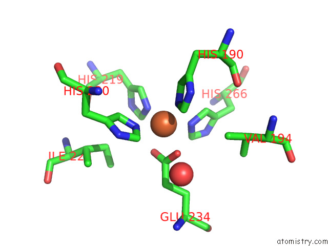
Mono view
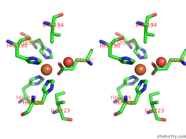
Stereo pair view
Mono view
Stereo pair view
A full contact list of Iron with other atoms in the Fe binding
site number 1 of Crystal Structure of R. Sphaeroides Photosynthetic Reaction Center Variant; W252V Mutant within 5.0Å range:
|
Reference:
J.B.Weaver,
C.Y.Lin,
K.M.Faries,
I.I.Mathews,
S.Russi,
D.Holten,
C.Kirmaier,
S.G.Boxer.
Photosynthetic Reaction Center Variants Made Via Genetic Code Expansion Indicate Tyrosine at M210 Tunes the Mechanism For Primary Electron Transfer Thesis pH.D. Stanford 294 2022UNIVERSITY.
Page generated: Thu Aug 8 09:16:25 2024
Last articles
Zn in 9J0NZn in 9J0O
Zn in 9J0P
Zn in 9FJX
Zn in 9EKB
Zn in 9C0F
Zn in 9CAH
Zn in 9CH0
Zn in 9CH3
Zn in 9CH1