Iron »
PDB 7mh9-7ni1 »
7nem »
Iron in PDB 7nem: Hydrogenase-2 Variant R479K - Anaerobically Oxidised Form
Enzymatic activity of Hydrogenase-2 Variant R479K - Anaerobically Oxidised Form
All present enzymatic activity of Hydrogenase-2 Variant R479K - Anaerobically Oxidised Form:
1.12.99.6;
1.12.99.6;
Protein crystallography data
The structure of Hydrogenase-2 Variant R479K - Anaerobically Oxidised Form, PDB code: 7nem
was solved by
S.B.Carr,
with X-Ray Crystallography technique. A brief refinement statistics is given in the table below:
| Resolution Low / High (Å) | 44.80 / 1.35 |
| Space group | P 21 21 21 |
| Cell size a, b, c (Å), α, β, γ (°) | 99.402, 100.252, 168.536, 90, 90, 90 |
| R / Rfree (%) | 15 / 16.7 |
Other elements in 7nem:
The structure of Hydrogenase-2 Variant R479K - Anaerobically Oxidised Form also contains other interesting chemical elements:
| Magnesium | (Mg) | 4 atoms |
| Nickel | (Ni) | 2 atoms |
| Chlorine | (Cl) | 1 atom |
Iron Binding Sites:
Pages:
>>> Page 1 <<< Page 2, Binding sites: 11 - 20; Page 3, Binding sites: 21 - 24;Binding sites:
The binding sites of Iron atom in the Hydrogenase-2 Variant R479K - Anaerobically Oxidised Form (pdb code 7nem). This binding sites where shown within 5.0 Angstroms radius around Iron atom.In total 24 binding sites of Iron where determined in the Hydrogenase-2 Variant R479K - Anaerobically Oxidised Form, PDB code: 7nem:
Jump to Iron binding site number: 1; 2; 3; 4; 5; 6; 7; 8; 9; 10;
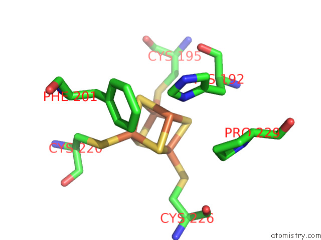
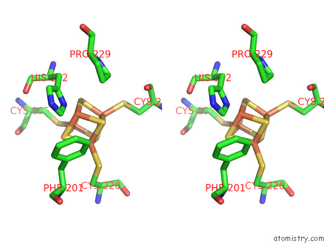
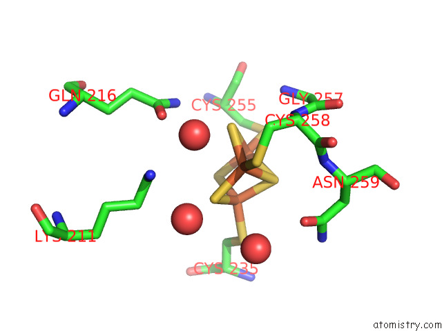
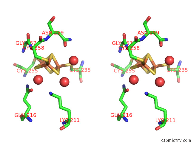
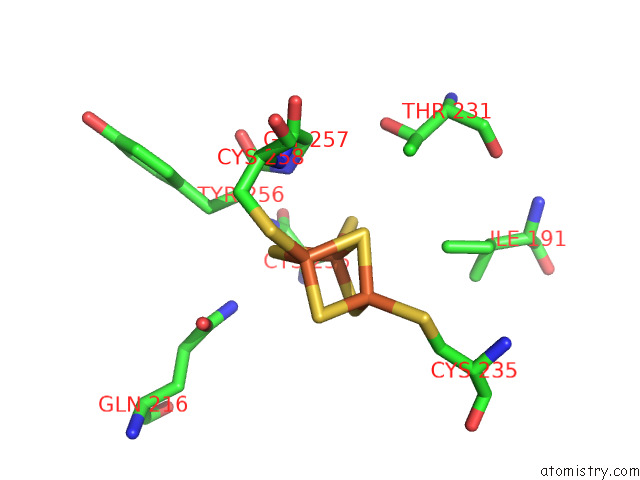
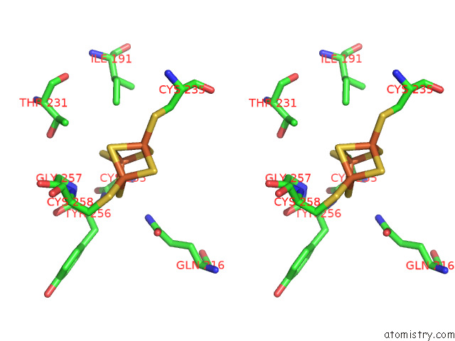
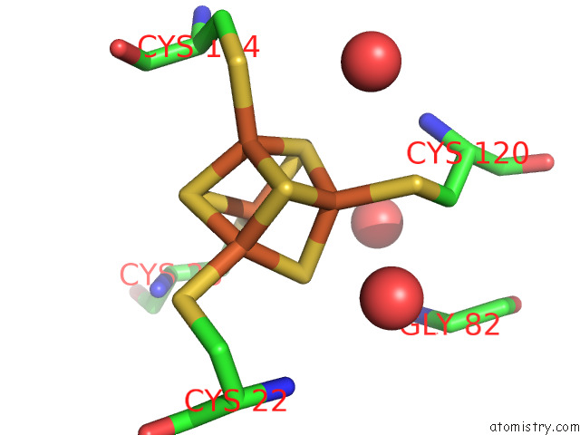
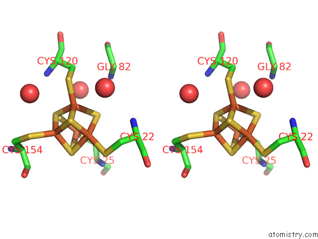
Iron binding site 1 out of 24 in 7nem
Go back to
Iron binding site 1 out
of 24 in the Hydrogenase-2 Variant R479K - Anaerobically Oxidised Form
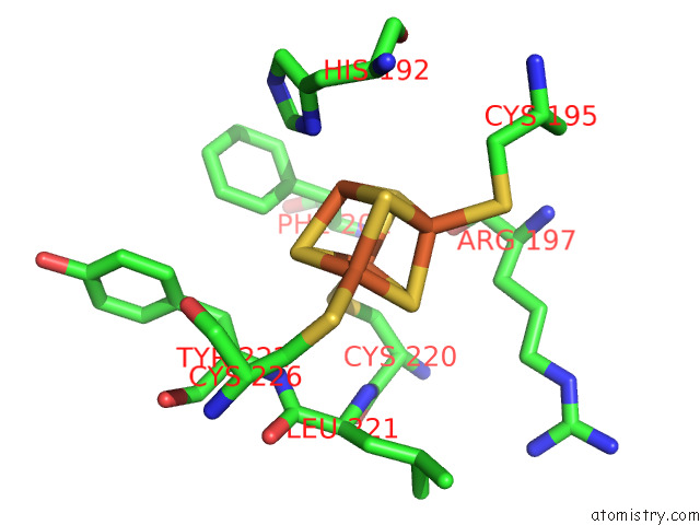
Mono view
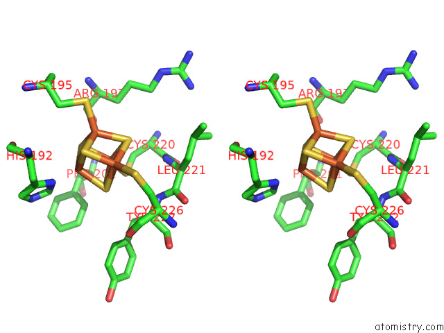
Stereo pair view
Mono view
Stereo pair view
A full contact list of Iron with other atoms in the Fe binding
site number 1 of Hydrogenase-2 Variant R479K - Anaerobically Oxidised Form within 5.0Å range:
|
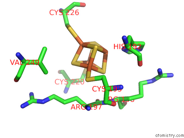
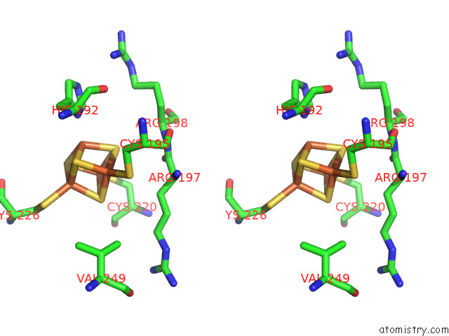
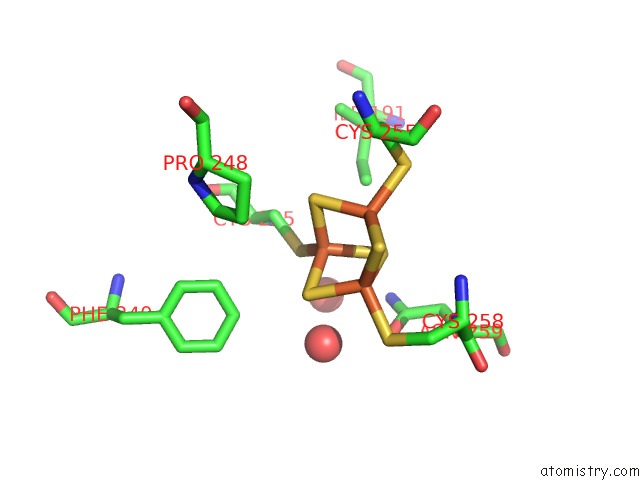
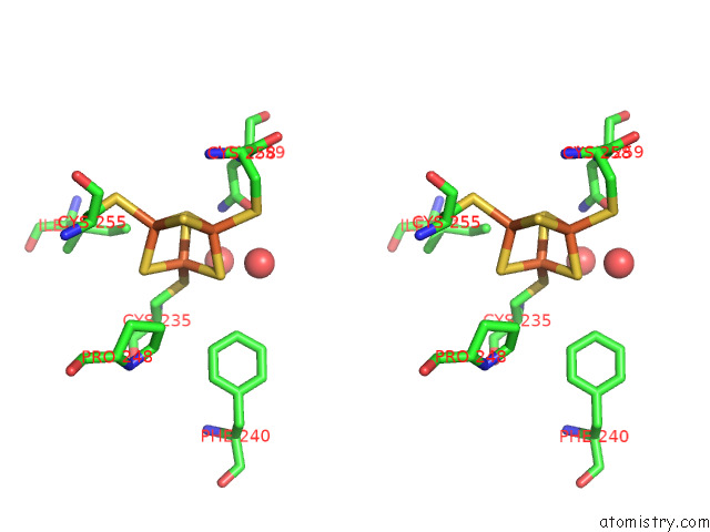
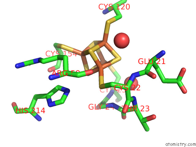
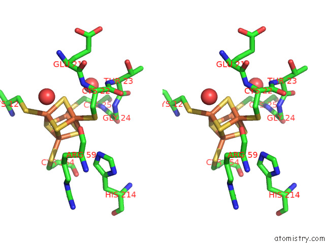
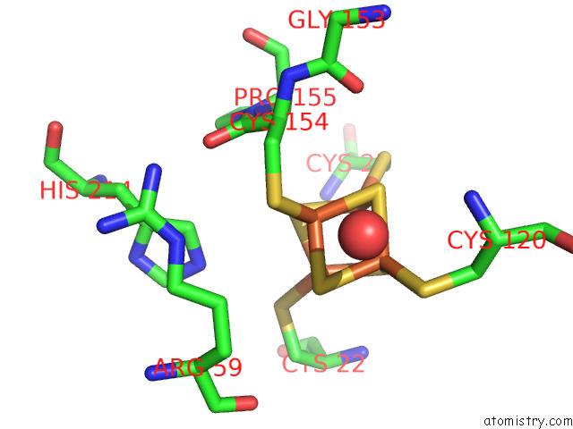
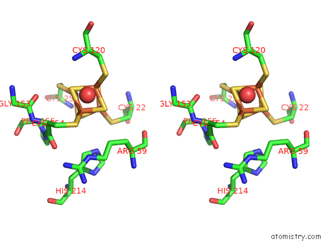
Iron binding site 2 out of 24 in 7nem
Go back to
Iron binding site 2 out
of 24 in the Hydrogenase-2 Variant R479K - Anaerobically Oxidised Form
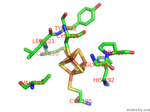
Mono view
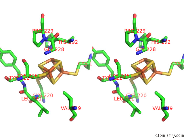
Stereo pair view
Mono view
Stereo pair view
A full contact list of Iron with other atoms in the Fe binding
site number 2 of Hydrogenase-2 Variant R479K - Anaerobically Oxidised Form within 5.0Å range:
|
Iron binding site 3 out of 24 in 7nem
Go back to
Iron binding site 3 out
of 24 in the Hydrogenase-2 Variant R479K - Anaerobically Oxidised Form
Mono view
Stereo pair view
Mono view
Stereo pair view
A full contact list of Iron with other atoms in the Fe binding
site number 3 of Hydrogenase-2 Variant R479K - Anaerobically Oxidised Form within 5.0Å range:
|
Iron binding site 4 out of 24 in 7nem
Go back to
Iron binding site 4 out
of 24 in the Hydrogenase-2 Variant R479K - Anaerobically Oxidised Form
Mono view
Stereo pair view
Mono view
Stereo pair view
A full contact list of Iron with other atoms in the Fe binding
site number 4 of Hydrogenase-2 Variant R479K - Anaerobically Oxidised Form within 5.0Å range:
|
Iron binding site 5 out of 24 in 7nem
Go back to
Iron binding site 5 out
of 24 in the Hydrogenase-2 Variant R479K - Anaerobically Oxidised Form
Mono view
Stereo pair view
Mono view
Stereo pair view
A full contact list of Iron with other atoms in the Fe binding
site number 5 of Hydrogenase-2 Variant R479K - Anaerobically Oxidised Form within 5.0Å range:
|
Iron binding site 6 out of 24 in 7nem
Go back to
Iron binding site 6 out
of 24 in the Hydrogenase-2 Variant R479K - Anaerobically Oxidised Form
Mono view
Stereo pair view
Mono view
Stereo pair view
A full contact list of Iron with other atoms in the Fe binding
site number 6 of Hydrogenase-2 Variant R479K - Anaerobically Oxidised Form within 5.0Å range:
|
Iron binding site 7 out of 24 in 7nem
Go back to
Iron binding site 7 out
of 24 in the Hydrogenase-2 Variant R479K - Anaerobically Oxidised Form
Mono view
Stereo pair view
Mono view
Stereo pair view
A full contact list of Iron with other atoms in the Fe binding
site number 7 of Hydrogenase-2 Variant R479K - Anaerobically Oxidised Form within 5.0Å range:
|
Iron binding site 8 out of 24 in 7nem
Go back to
Iron binding site 8 out
of 24 in the Hydrogenase-2 Variant R479K - Anaerobically Oxidised Form
Mono view
Stereo pair view
Mono view
Stereo pair view
A full contact list of Iron with other atoms in the Fe binding
site number 8 of Hydrogenase-2 Variant R479K - Anaerobically Oxidised Form within 5.0Å range:
|
Iron binding site 9 out of 24 in 7nem
Go back to
Iron binding site 9 out
of 24 in the Hydrogenase-2 Variant R479K - Anaerobically Oxidised Form
Mono view
Stereo pair view
Mono view
Stereo pair view
A full contact list of Iron with other atoms in the Fe binding
site number 9 of Hydrogenase-2 Variant R479K - Anaerobically Oxidised Form within 5.0Å range:
|
Iron binding site 10 out of 24 in 7nem
Go back to
Iron binding site 10 out
of 24 in the Hydrogenase-2 Variant R479K - Anaerobically Oxidised Form
Mono view
Stereo pair view
Mono view
Stereo pair view
A full contact list of Iron with other atoms in the Fe binding
site number 10 of Hydrogenase-2 Variant R479K - Anaerobically Oxidised Form within 5.0Å range:
|
Reference:
R.M.Evans,
S.E.Beaton,
L.Kertiss,
W.K.Myers,
S.B.Carr,
F.A.Armstrong.
A Comprehensive Structural and Kinetic Investigation of the Role of the Active-Site Argininein Bidirectional Hydrogen Activation By the [Nife]-Hydrogenase "Hyd-2) From Escherichia Coli To Be Published.
Page generated: Thu Aug 8 09:33:23 2024
Last articles
Fe in 2YXOFe in 2YRS
Fe in 2YXC
Fe in 2YNM
Fe in 2YVJ
Fe in 2YP1
Fe in 2YU2
Fe in 2YU1
Fe in 2YQB
Fe in 2YOO