Iron in PDB 7nkz: Cryo-Em Structure of the Cytochrome Bd Oxidase From M. Tuberculosis at 2.5 A Resolution
Iron Binding Sites:
The binding sites of Iron atom in the Cryo-Em Structure of the Cytochrome Bd Oxidase From M. Tuberculosis at 2.5 A Resolution
(pdb code 7nkz). This binding sites where shown within
5.0 Angstroms radius around Iron atom.
In total 3 binding sites of Iron where determined in the
Cryo-Em Structure of the Cytochrome Bd Oxidase From M. Tuberculosis at 2.5 A Resolution, PDB code: 7nkz:
Jump to Iron binding site number:
1;
2;
3;
Iron binding site 1 out
of 3 in 7nkz
Go back to
Iron Binding Sites List in 7nkz
Iron binding site 1 out
of 3 in the Cryo-Em Structure of the Cytochrome Bd Oxidase From M. Tuberculosis at 2.5 A Resolution
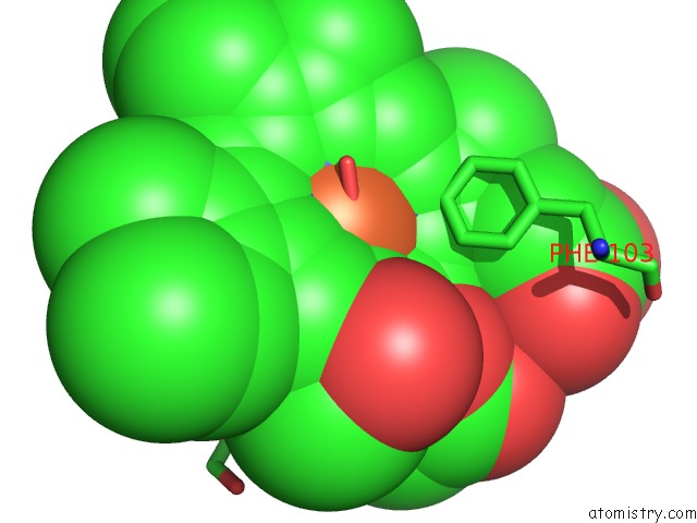
Mono view
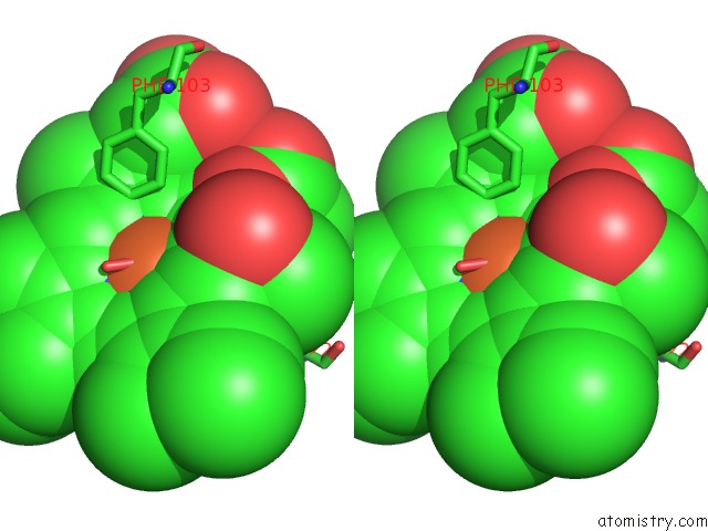
Stereo pair view
|
A full contact list of Iron with other atoms in the Fe binding
site number 1 of Cryo-Em Structure of the Cytochrome Bd Oxidase From M. Tuberculosis at 2.5 A Resolution within 5.0Å range:
probe
|
atom
|
residue
|
distance (Å)
|
B
|
Occ
|
A:Fe703
b:33.3
occ:1.00
|
FE
|
A:HDD703
|
0.0
|
33.3
|
1.0
|
ND
|
A:HDD703
|
1.8
|
33.3
|
1.0
|
NA
|
A:HDD703
|
2.1
|
33.3
|
1.0
|
NC
|
A:HDD703
|
2.1
|
33.3
|
1.0
|
NB
|
A:HDD703
|
2.1
|
33.3
|
1.0
|
NE2
|
A:HIS18
|
2.7
|
29.4
|
1.0
|
C4D
|
A:HDD703
|
2.8
|
33.3
|
1.0
|
C1D
|
A:HDD703
|
2.8
|
33.3
|
1.0
|
C4C
|
A:HDD703
|
3.0
|
33.3
|
1.0
|
C1A
|
A:HDD703
|
3.0
|
33.3
|
1.0
|
C1B
|
A:HDD703
|
3.1
|
33.3
|
1.0
|
C4B
|
A:HDD703
|
3.1
|
33.3
|
1.0
|
C4A
|
A:HDD703
|
3.1
|
33.3
|
1.0
|
C1C
|
A:HDD703
|
3.1
|
33.3
|
1.0
|
CHA
|
A:HDD703
|
3.3
|
33.3
|
1.0
|
CHD
|
A:HDD703
|
3.3
|
33.3
|
1.0
|
O1
|
A:OXY701
|
3.3
|
38.1
|
1.0
|
CHC
|
A:HDD703
|
3.5
|
33.3
|
1.0
|
CHB
|
A:HDD703
|
3.5
|
33.3
|
1.0
|
CE1
|
A:HIS18
|
3.6
|
29.4
|
1.0
|
CD2
|
A:HIS18
|
3.6
|
29.4
|
1.0
|
O2
|
A:OXY701
|
3.9
|
38.1
|
1.0
|
C3D
|
A:HDD703
|
4.1
|
33.3
|
1.0
|
C2D
|
A:HDD703
|
4.1
|
33.3
|
1.0
|
C3C
|
A:HDD703
|
4.3
|
33.3
|
1.0
|
C2A
|
A:HDD703
|
4.4
|
33.3
|
1.0
|
CZ
|
A:PHE103
|
4.4
|
38.1
|
1.0
|
C2B
|
A:HDD703
|
4.4
|
33.3
|
1.0
|
C3B
|
A:HDD703
|
4.4
|
33.3
|
1.0
|
C3A
|
A:HDD703
|
4.4
|
33.3
|
1.0
|
C2C
|
A:HDD703
|
4.4
|
33.3
|
1.0
|
CG2
|
A:VAL22
|
4.6
|
29.6
|
1.0
|
ND1
|
A:HIS18
|
4.7
|
29.4
|
1.0
|
OND
|
A:HDD703
|
4.8
|
33.3
|
1.0
|
CG
|
A:HIS18
|
4.8
|
29.4
|
1.0
|
CE1
|
A:PHE103
|
4.8
|
38.1
|
1.0
|
CAD
|
A:HDD703
|
4.9
|
33.3
|
1.0
|
O1D
|
A:HDD703
|
5.0
|
33.3
|
1.0
|
|
Iron binding site 2 out
of 3 in 7nkz
Go back to
Iron Binding Sites List in 7nkz
Iron binding site 2 out
of 3 in the Cryo-Em Structure of the Cytochrome Bd Oxidase From M. Tuberculosis at 2.5 A Resolution
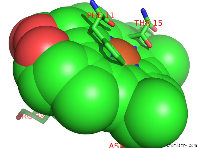
Mono view
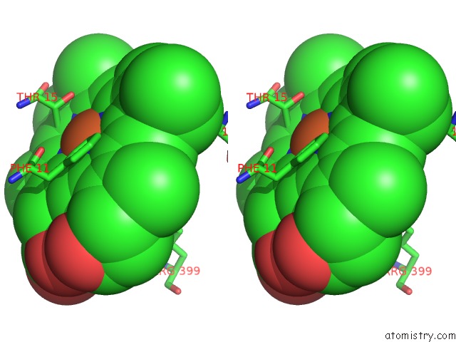
Stereo pair view
|
A full contact list of Iron with other atoms in the Fe binding
site number 2 of Cryo-Em Structure of the Cytochrome Bd Oxidase From M. Tuberculosis at 2.5 A Resolution within 5.0Å range:
probe
|
atom
|
residue
|
distance (Å)
|
B
|
Occ
|
A:Fe704
b:36.1
occ:1.00
|
FE
|
A:HEB704
|
0.0
|
36.1
|
1.0
|
NA
|
A:HEB704
|
2.1
|
36.1
|
1.0
|
NB
|
A:HEB704
|
2.2
|
36.1
|
1.0
|
NC
|
A:HEB704
|
2.2
|
36.1
|
1.0
|
ND
|
A:HEB704
|
2.3
|
36.1
|
1.0
|
OE2
|
A:GLU396
|
2.7
|
37.1
|
1.0
|
C1A
|
A:HEB704
|
3.2
|
36.1
|
1.0
|
C1C
|
A:HEB704
|
3.2
|
36.1
|
1.0
|
C1B
|
A:HEB704
|
3.3
|
36.1
|
1.0
|
C4A
|
A:HEB704
|
3.3
|
36.1
|
1.0
|
C4B
|
A:HEB704
|
3.3
|
36.1
|
1.0
|
C4C
|
A:HEB704
|
3.3
|
36.1
|
1.0
|
C4D
|
A:HEB704
|
3.4
|
36.1
|
1.0
|
C1D
|
A:HEB704
|
3.4
|
36.1
|
1.0
|
CD
|
A:GLU396
|
3.5
|
37.1
|
1.0
|
CG
|
A:GLU396
|
3.6
|
37.1
|
1.0
|
CHC
|
A:HEB704
|
3.7
|
36.1
|
1.0
|
CE1
|
A:PHE11
|
3.7
|
35.9
|
1.0
|
CHB
|
A:HEB704
|
3.7
|
36.1
|
1.0
|
CHA
|
A:HEB704
|
3.7
|
36.1
|
1.0
|
CHD
|
A:HEB704
|
3.8
|
36.1
|
1.0
|
CD1
|
A:PHE11
|
4.0
|
35.9
|
1.0
|
CG2
|
A:THR15
|
4.2
|
33.0
|
1.0
|
C2A
|
A:HEB704
|
4.2
|
36.1
|
1.0
|
C2C
|
A:HEB704
|
4.2
|
36.1
|
1.0
|
C2B
|
A:HEB704
|
4.2
|
36.1
|
1.0
|
C3A
|
A:HEB704
|
4.3
|
36.1
|
1.0
|
C3B
|
A:HEB704
|
4.3
|
36.1
|
1.0
|
C3C
|
A:HEB704
|
4.4
|
36.1
|
1.0
|
C2D
|
A:HEB704
|
4.4
|
36.1
|
1.0
|
C3D
|
A:HEB704
|
4.6
|
36.1
|
1.0
|
OE1
|
A:GLU396
|
4.6
|
37.1
|
1.0
|
CB
|
A:GLU396
|
4.6
|
37.1
|
1.0
|
NH2
|
A:ARG399
|
4.6
|
37.2
|
1.0
|
CZ
|
A:PHE11
|
4.8
|
35.9
|
1.0
|
ND2
|
A:ASN147
|
4.9
|
34.3
|
1.0
|
OG1
|
A:THR15
|
4.9
|
33.0
|
1.0
|
|
Iron binding site 3 out
of 3 in 7nkz
Go back to
Iron Binding Sites List in 7nkz
Iron binding site 3 out
of 3 in the Cryo-Em Structure of the Cytochrome Bd Oxidase From M. Tuberculosis at 2.5 A Resolution
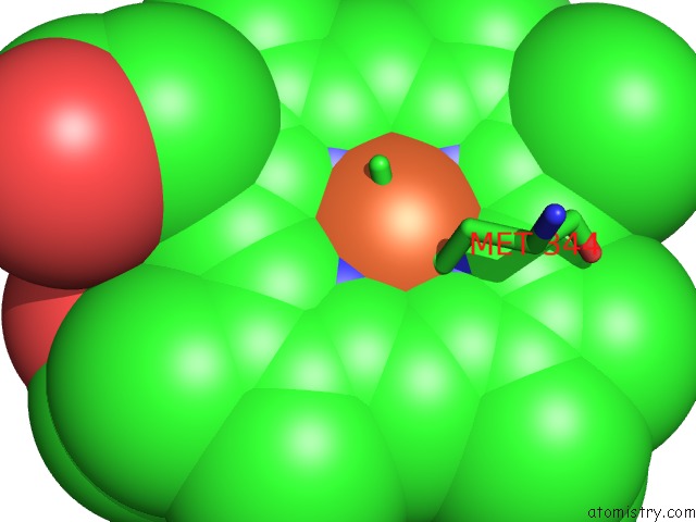
Mono view
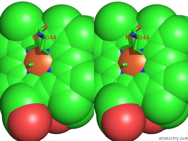
Stereo pair view
|
A full contact list of Iron with other atoms in the Fe binding
site number 3 of Cryo-Em Structure of the Cytochrome Bd Oxidase From M. Tuberculosis at 2.5 A Resolution within 5.0Å range:
probe
|
atom
|
residue
|
distance (Å)
|
B
|
Occ
|
A:Fe705
b:40.2
occ:1.00
|
FE
|
A:HEB705
|
0.0
|
40.2
|
1.0
|
NA
|
A:HEB705
|
2.1
|
40.2
|
1.0
|
NB
|
A:HEB705
|
2.2
|
40.2
|
1.0
|
NC
|
A:HEB705
|
2.2
|
40.2
|
1.0
|
ND
|
A:HEB705
|
2.3
|
40.2
|
1.0
|
SD
|
A:MET344
|
2.3
|
44.3
|
1.0
|
NE2
|
A:HIS185
|
2.6
|
36.4
|
1.0
|
C1A
|
A:HEB705
|
3.2
|
40.2
|
1.0
|
C1C
|
A:HEB705
|
3.2
|
40.2
|
1.0
|
C1B
|
A:HEB705
|
3.3
|
40.2
|
1.0
|
C4A
|
A:HEB705
|
3.3
|
40.2
|
1.0
|
C4B
|
A:HEB705
|
3.3
|
40.2
|
1.0
|
C4C
|
A:HEB705
|
3.3
|
40.2
|
1.0
|
C4D
|
A:HEB705
|
3.4
|
40.2
|
1.0
|
C1D
|
A:HEB705
|
3.4
|
40.2
|
1.0
|
CD2
|
A:HIS185
|
3.4
|
36.4
|
1.0
|
CE1
|
A:HIS185
|
3.6
|
36.4
|
1.0
|
CG
|
A:MET344
|
3.6
|
44.3
|
1.0
|
CHB
|
A:HEB705
|
3.7
|
40.2
|
1.0
|
CHC
|
A:HEB705
|
3.7
|
40.2
|
1.0
|
CHA
|
A:HEB705
|
3.7
|
40.2
|
1.0
|
CHD
|
A:HEB705
|
3.8
|
40.2
|
1.0
|
CE
|
A:MET344
|
3.8
|
44.3
|
1.0
|
C2A
|
A:HEB705
|
4.2
|
40.2
|
1.0
|
C2C
|
A:HEB705
|
4.2
|
40.2
|
1.0
|
C2B
|
A:HEB705
|
4.2
|
40.2
|
1.0
|
C3A
|
A:HEB705
|
4.3
|
40.2
|
1.0
|
C3B
|
A:HEB705
|
4.3
|
40.2
|
1.0
|
C3C
|
A:HEB705
|
4.4
|
40.2
|
1.0
|
C2D
|
A:HEB705
|
4.4
|
40.2
|
1.0
|
C3D
|
A:HEB705
|
4.6
|
40.2
|
1.0
|
CG
|
A:HIS185
|
4.6
|
36.4
|
1.0
|
ND1
|
A:HIS185
|
4.7
|
36.4
|
1.0
|
CB
|
A:MET344
|
4.9
|
44.3
|
1.0
|
|
Reference:
S.Safarian,
H.K.Opel-Reading,
D.Wu,
A.R.Mehdipour,
K.Hards,
L.K.Harold,
M.Radloff,
I.Stewart,
S.Welsch,
G.Hummer,
G.M.Cook,
K.L.Krause,
H.Michel.
The Cryo-Em Structure of the Bd Oxidase From M. Tuberculosis Reveals A Unique Structural Framework and Enables Rational Drug Design to Combat Tb Nat Commun 2021.
ISSN: ESSN 2041-1723
DOI: 10.1038/S41467-021-25537-Z
Page generated: Thu Aug 8 09:50:36 2024
|