Iron »
PDB 7o4j-7oqr »
7o6c »
Iron in PDB 7o6c: Crystal Structure of Human Mitochondrial Ferritin (Hmtf) Fe(II)-Loaded For 15 Minutes Under Anaerobic Environment
Enzymatic activity of Crystal Structure of Human Mitochondrial Ferritin (Hmtf) Fe(II)-Loaded For 15 Minutes Under Anaerobic Environment
All present enzymatic activity of Crystal Structure of Human Mitochondrial Ferritin (Hmtf) Fe(II)-Loaded For 15 Minutes Under Anaerobic Environment:
1.16.3.1;
1.16.3.1;
Protein crystallography data
The structure of Crystal Structure of Human Mitochondrial Ferritin (Hmtf) Fe(II)-Loaded For 15 Minutes Under Anaerobic Environment, PDB code: 7o6c
was solved by
C.Pozzi,
S.Ciambellotti,
G.Tassone,
P.Turano,
S.Mangani,
with X-Ray Crystallography technique. A brief refinement statistics is given in the table below:
| Resolution Low / High (Å) | 52.72 / 1.20 |
| Space group | F 4 3 2 |
| Cell size a, b, c (Å), α, β, γ (°) | 182.449, 182.449, 182.449, 90, 90, 90 |
| R / Rfree (%) | 12.1 / 14.3 |
Other elements in 7o6c:
The structure of Crystal Structure of Human Mitochondrial Ferritin (Hmtf) Fe(II)-Loaded For 15 Minutes Under Anaerobic Environment also contains other interesting chemical elements:
| Magnesium | (Mg) | 6 atoms |
| Chlorine | (Cl) | 11 atoms |
Iron Binding Sites:
The binding sites of Iron atom in the Crystal Structure of Human Mitochondrial Ferritin (Hmtf) Fe(II)-Loaded For 15 Minutes Under Anaerobic Environment
(pdb code 7o6c). This binding sites where shown within
5.0 Angstroms radius around Iron atom.
In total 8 binding sites of Iron where determined in the Crystal Structure of Human Mitochondrial Ferritin (Hmtf) Fe(II)-Loaded For 15 Minutes Under Anaerobic Environment, PDB code: 7o6c:
Jump to Iron binding site number: 1; 2; 3; 4; 5; 6; 7; 8;
In total 8 binding sites of Iron where determined in the Crystal Structure of Human Mitochondrial Ferritin (Hmtf) Fe(II)-Loaded For 15 Minutes Under Anaerobic Environment, PDB code: 7o6c:
Jump to Iron binding site number: 1; 2; 3; 4; 5; 6; 7; 8;
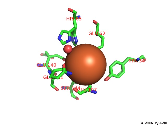
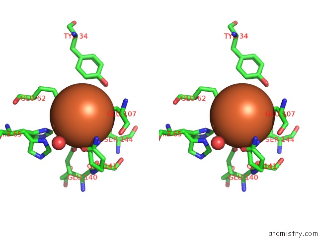
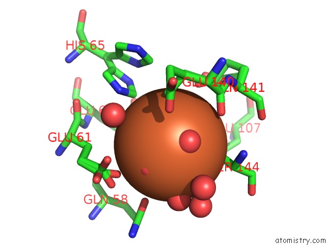
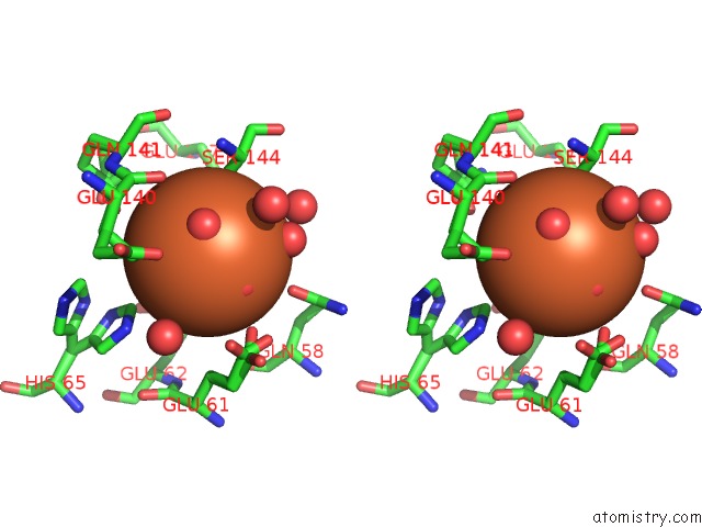
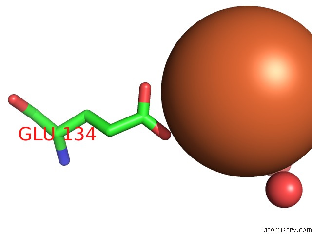
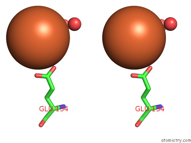
Iron binding site 1 out of 8 in 7o6c
Go back to
Iron binding site 1 out
of 8 in the Crystal Structure of Human Mitochondrial Ferritin (Hmtf) Fe(II)-Loaded For 15 Minutes Under Anaerobic Environment
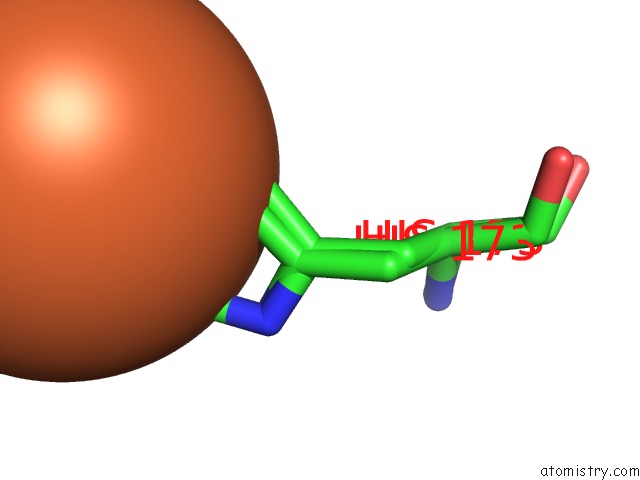
Mono view
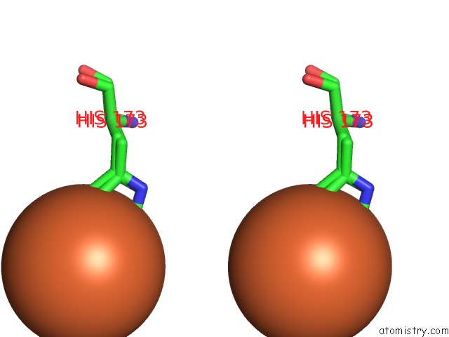
Stereo pair view
Mono view
Stereo pair view
A full contact list of Iron with other atoms in the Fe binding
site number 1 of Crystal Structure of Human Mitochondrial Ferritin (Hmtf) Fe(II)-Loaded For 15 Minutes Under Anaerobic Environment within 5.0Å range:
|
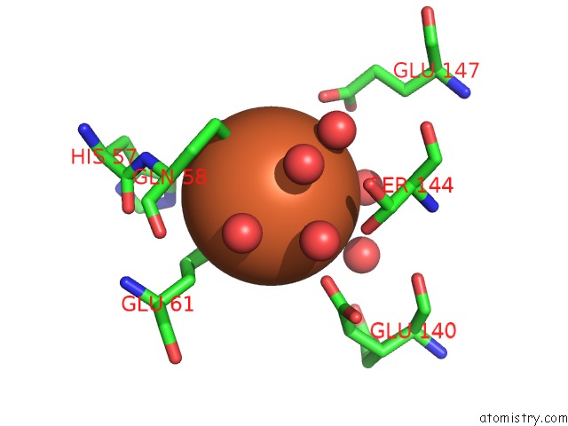
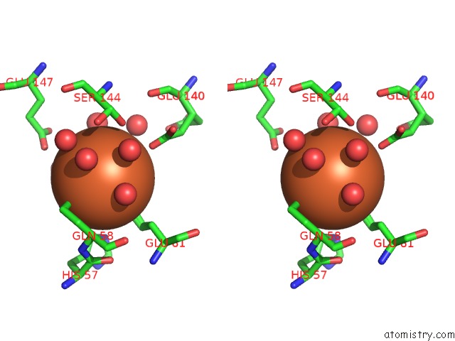
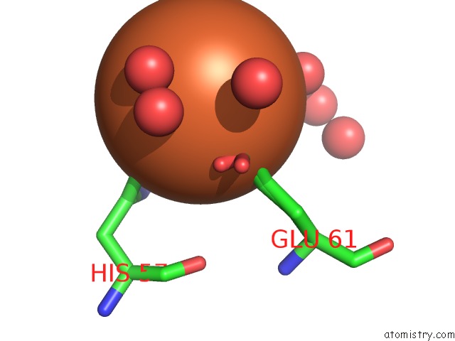
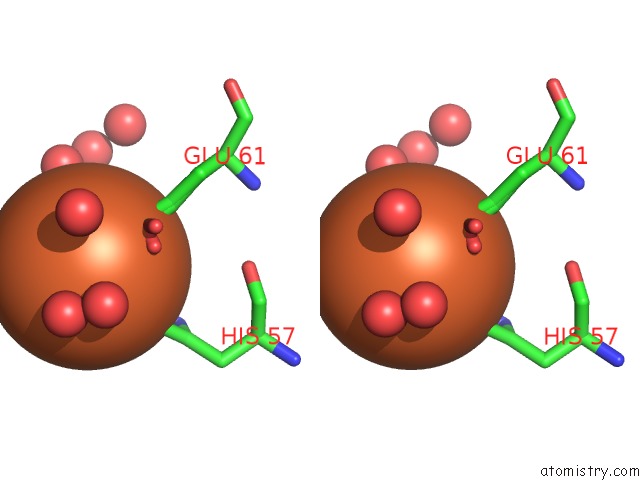
Iron binding site 2 out of 8 in 7o6c
Go back to
Iron binding site 2 out
of 8 in the Crystal Structure of Human Mitochondrial Ferritin (Hmtf) Fe(II)-Loaded For 15 Minutes Under Anaerobic Environment
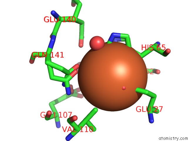
Mono view
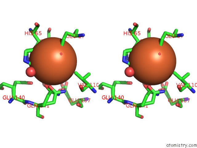
Stereo pair view
Mono view
Stereo pair view
A full contact list of Iron with other atoms in the Fe binding
site number 2 of Crystal Structure of Human Mitochondrial Ferritin (Hmtf) Fe(II)-Loaded For 15 Minutes Under Anaerobic Environment within 5.0Å range:
|
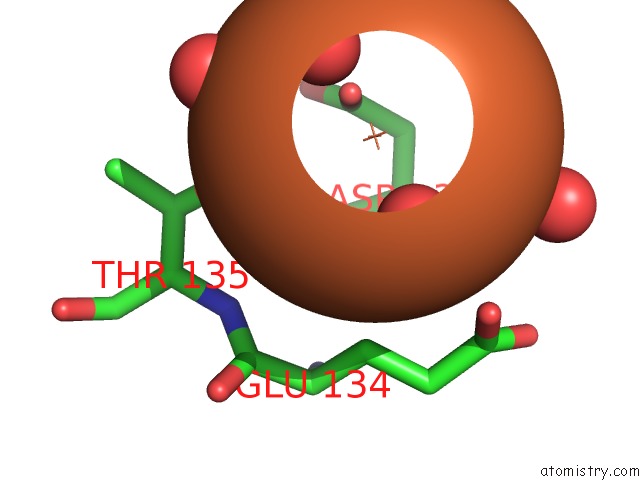
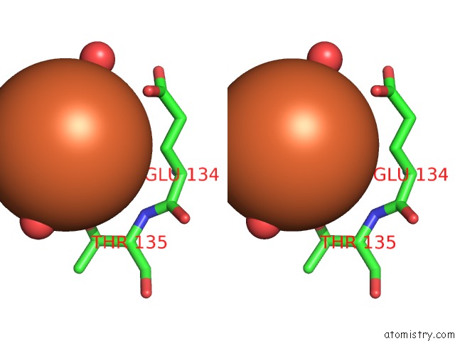
Iron binding site 3 out of 8 in 7o6c
Go back to
Iron binding site 3 out
of 8 in the Crystal Structure of Human Mitochondrial Ferritin (Hmtf) Fe(II)-Loaded For 15 Minutes Under Anaerobic Environment
Mono view
Stereo pair view
Mono view
Stereo pair view
A full contact list of Iron with other atoms in the Fe binding
site number 3 of Crystal Structure of Human Mitochondrial Ferritin (Hmtf) Fe(II)-Loaded For 15 Minutes Under Anaerobic Environment within 5.0Å range:
|
Iron binding site 4 out of 8 in 7o6c
Go back to
Iron binding site 4 out
of 8 in the Crystal Structure of Human Mitochondrial Ferritin (Hmtf) Fe(II)-Loaded For 15 Minutes Under Anaerobic Environment
Mono view
Stereo pair view
Mono view
Stereo pair view
A full contact list of Iron with other atoms in the Fe binding
site number 4 of Crystal Structure of Human Mitochondrial Ferritin (Hmtf) Fe(II)-Loaded For 15 Minutes Under Anaerobic Environment within 5.0Å range:
|
Iron binding site 5 out of 8 in 7o6c
Go back to
Iron binding site 5 out
of 8 in the Crystal Structure of Human Mitochondrial Ferritin (Hmtf) Fe(II)-Loaded For 15 Minutes Under Anaerobic Environment
Mono view
Stereo pair view
Mono view
Stereo pair view
A full contact list of Iron with other atoms in the Fe binding
site number 5 of Crystal Structure of Human Mitochondrial Ferritin (Hmtf) Fe(II)-Loaded For 15 Minutes Under Anaerobic Environment within 5.0Å range:
|
Iron binding site 6 out of 8 in 7o6c
Go back to
Iron binding site 6 out
of 8 in the Crystal Structure of Human Mitochondrial Ferritin (Hmtf) Fe(II)-Loaded For 15 Minutes Under Anaerobic Environment
Mono view
Stereo pair view
Mono view
Stereo pair view
A full contact list of Iron with other atoms in the Fe binding
site number 6 of Crystal Structure of Human Mitochondrial Ferritin (Hmtf) Fe(II)-Loaded For 15 Minutes Under Anaerobic Environment within 5.0Å range:
|
Iron binding site 7 out of 8 in 7o6c
Go back to
Iron binding site 7 out
of 8 in the Crystal Structure of Human Mitochondrial Ferritin (Hmtf) Fe(II)-Loaded For 15 Minutes Under Anaerobic Environment
Mono view
Stereo pair view
Mono view
Stereo pair view
A full contact list of Iron with other atoms in the Fe binding
site number 7 of Crystal Structure of Human Mitochondrial Ferritin (Hmtf) Fe(II)-Loaded For 15 Minutes Under Anaerobic Environment within 5.0Å range:
|
Iron binding site 8 out of 8 in 7o6c
Go back to
Iron binding site 8 out
of 8 in the Crystal Structure of Human Mitochondrial Ferritin (Hmtf) Fe(II)-Loaded For 15 Minutes Under Anaerobic Environment
Mono view
Stereo pair view
Mono view
Stereo pair view
A full contact list of Iron with other atoms in the Fe binding
site number 8 of Crystal Structure of Human Mitochondrial Ferritin (Hmtf) Fe(II)-Loaded For 15 Minutes Under Anaerobic Environment within 5.0Å range:
|
Reference:
S.Ciambellotti,
A.Pratesi,
G.Tassone,
P.Turano,
S.Mangani,
C.Pozzi.
Iron Binding in the Ferroxidase Site of Human Mitochondrial Ferritin. Chemistry 2021.
ISSN: ISSN 0947-6539
PubMed: 34343376
DOI: 10.1002/CHEM.202102270
Page generated: Thu Aug 8 14:06:32 2024
ISSN: ISSN 0947-6539
PubMed: 34343376
DOI: 10.1002/CHEM.202102270
Last articles
Zn in 9J0NZn in 9J0O
Zn in 9J0P
Zn in 9FJX
Zn in 9EKB
Zn in 9C0F
Zn in 9CAH
Zn in 9CH0
Zn in 9CH3
Zn in 9CH1