Iron »
PDB 7o4j-7oqr »
7o8d »
Iron in PDB 7o8d: Dife-Sulerythrin Oxidised By H2O2
Protein crystallography data
The structure of Dife-Sulerythrin Oxidised By H2O2, PDB code: 7o8d
was solved by
J.-H.Jeoung,
H.Dobbek,
with X-Ray Crystallography technique. A brief refinement statistics is given in the table below:
| Resolution Low / High (Å) | 38.96 / 1.12 |
| Space group | P 63 |
| Cell size a, b, c (Å), α, β, γ (°) | 71.971, 71.971, 99.814, 90, 90, 120 |
| R / Rfree (%) | 13.7 / 16.9 |
Other elements in 7o8d:
The structure of Dife-Sulerythrin Oxidised By H2O2 also contains other interesting chemical elements:
| Chlorine | (Cl) | 2 atoms |
Iron Binding Sites:
The binding sites of Iron atom in the Dife-Sulerythrin Oxidised By H2O2
(pdb code 7o8d). This binding sites where shown within
5.0 Angstroms radius around Iron atom.
In total 4 binding sites of Iron where determined in the Dife-Sulerythrin Oxidised By H2O2, PDB code: 7o8d:
Jump to Iron binding site number: 1; 2; 3; 4;
In total 4 binding sites of Iron where determined in the Dife-Sulerythrin Oxidised By H2O2, PDB code: 7o8d:
Jump to Iron binding site number: 1; 2; 3; 4;
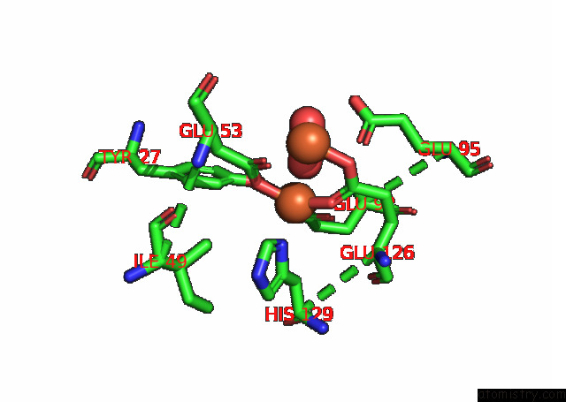
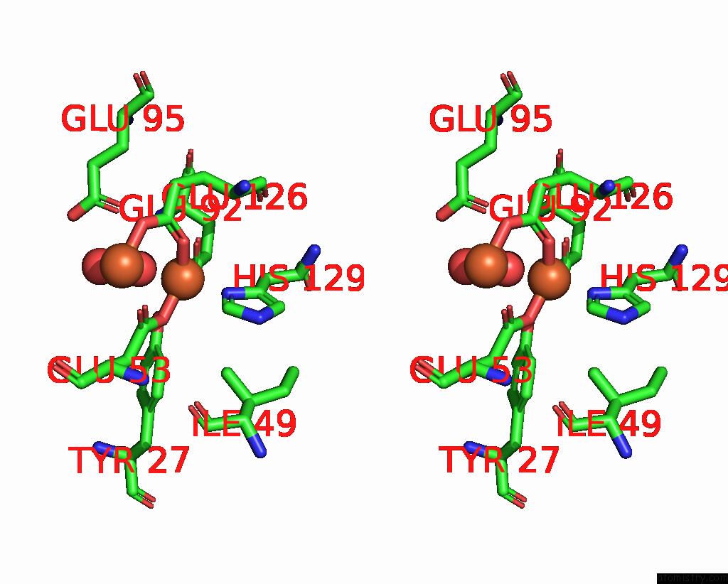
Iron binding site 1 out of 4 in 7o8d
Go back to
Iron binding site 1 out
of 4 in the Dife-Sulerythrin Oxidised By H2O2
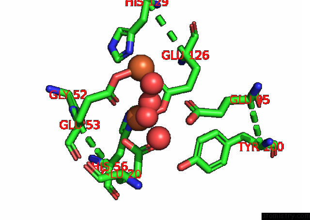
Mono view
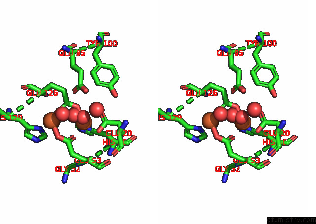
Stereo pair view
Mono view
Stereo pair view
A full contact list of Iron with other atoms in the Fe binding
site number 1 of Dife-Sulerythrin Oxidised By H2O2 within 5.0Å range:
|
Iron binding site 2 out of 4 in 7o8d
Go back to
Iron binding site 2 out
of 4 in the Dife-Sulerythrin Oxidised By H2O2
Mono view
Stereo pair view
Mono view
Stereo pair view
A full contact list of Iron with other atoms in the Fe binding
site number 2 of Dife-Sulerythrin Oxidised By H2O2 within 5.0Å range:
|
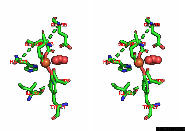
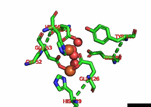
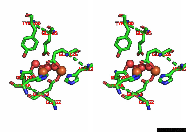
Iron binding site 3 out of 4 in 7o8d
Go back to
Iron binding site 3 out
of 4 in the Dife-Sulerythrin Oxidised By H2O2
Mono view
Stereo pair view
Mono view
Stereo pair view
A full contact list of Iron with other atoms in the Fe binding
site number 3 of Dife-Sulerythrin Oxidised By H2O2 within 5.0Å range:
|
Iron binding site 4 out of 4 in 7o8d
Go back to
Iron binding site 4 out
of 4 in the Dife-Sulerythrin Oxidised By H2O2
Mono view
Stereo pair view
Mono view
Stereo pair view
A full contact list of Iron with other atoms in the Fe binding
site number 4 of Dife-Sulerythrin Oxidised By H2O2 within 5.0Å range:
|
Reference:
J.H.Jeoung,
S.Runger,
M.Haumann,
B.Neumann,
F.Klemke,
V.Davis,
A.Fischer,
H.Dau,
U.Wollenberger,
H.Dobbek.
Bimetallic Mn, Fe, Co, and Ni Sites in A Four-Helix Bundle Protein: Metal Binding, Structure, and Peroxide Activation. Inorg.Chem. 2021.
ISSN: ISSN 0020-1669
PubMed: 34757735
DOI: 10.1021/ACS.INORGCHEM.1C01919
Page generated: Thu Aug 8 14:15:49 2024
ISSN: ISSN 0020-1669
PubMed: 34757735
DOI: 10.1021/ACS.INORGCHEM.1C01919
Last articles
Zn in 9J0NZn in 9J0O
Zn in 9J0P
Zn in 9FJX
Zn in 9EKB
Zn in 9C0F
Zn in 9CAH
Zn in 9CH0
Zn in 9CH3
Zn in 9CH1