Iron »
PDB 7oqy-7p7j »
7ov8 »
Iron in PDB 7ov8: Crystal Structure of Pig Purple Acid Phosphatase in Complex with 4-(2- Hydroxyethyl)-1-Piperazineethanesulfonic Acid (Hepes) and Glycerol
Enzymatic activity of Crystal Structure of Pig Purple Acid Phosphatase in Complex with 4-(2- Hydroxyethyl)-1-Piperazineethanesulfonic Acid (Hepes) and Glycerol
All present enzymatic activity of Crystal Structure of Pig Purple Acid Phosphatase in Complex with 4-(2- Hydroxyethyl)-1-Piperazineethanesulfonic Acid (Hepes) and Glycerol:
3.1.3.2;
3.1.3.2;
Protein crystallography data
The structure of Crystal Structure of Pig Purple Acid Phosphatase in Complex with 4-(2- Hydroxyethyl)-1-Piperazineethanesulfonic Acid (Hepes) and Glycerol, PDB code: 7ov8
was solved by
D.Feder,
R.P.Mcgeary,
L.W.Guddat,
G.Schenk,
with X-Ray Crystallography technique. A brief refinement statistics is given in the table below:
| Resolution Low / High (Å) | 39.57 / 2.30 |
| Space group | P 21 21 21 |
| Cell size a, b, c (Å), α, β, γ (°) | 62.382, 69.772, 75.309, 90, 90, 90 |
| R / Rfree (%) | 16.2 / 25.6 |
Other elements in 7ov8:
The structure of Crystal Structure of Pig Purple Acid Phosphatase in Complex with 4-(2- Hydroxyethyl)-1-Piperazineethanesulfonic Acid (Hepes) and Glycerol also contains other interesting chemical elements:
| Sodium | (Na) | 2 atoms |
Iron Binding Sites:
The binding sites of Iron atom in the Crystal Structure of Pig Purple Acid Phosphatase in Complex with 4-(2- Hydroxyethyl)-1-Piperazineethanesulfonic Acid (Hepes) and Glycerol
(pdb code 7ov8). This binding sites where shown within
5.0 Angstroms radius around Iron atom.
In total 2 binding sites of Iron where determined in the Crystal Structure of Pig Purple Acid Phosphatase in Complex with 4-(2- Hydroxyethyl)-1-Piperazineethanesulfonic Acid (Hepes) and Glycerol, PDB code: 7ov8:
Jump to Iron binding site number: 1; 2;
In total 2 binding sites of Iron where determined in the Crystal Structure of Pig Purple Acid Phosphatase in Complex with 4-(2- Hydroxyethyl)-1-Piperazineethanesulfonic Acid (Hepes) and Glycerol, PDB code: 7ov8:
Jump to Iron binding site number: 1; 2;
Iron binding site 1 out of 2 in 7ov8
Go back to
Iron binding site 1 out
of 2 in the Crystal Structure of Pig Purple Acid Phosphatase in Complex with 4-(2- Hydroxyethyl)-1-Piperazineethanesulfonic Acid (Hepes) and Glycerol
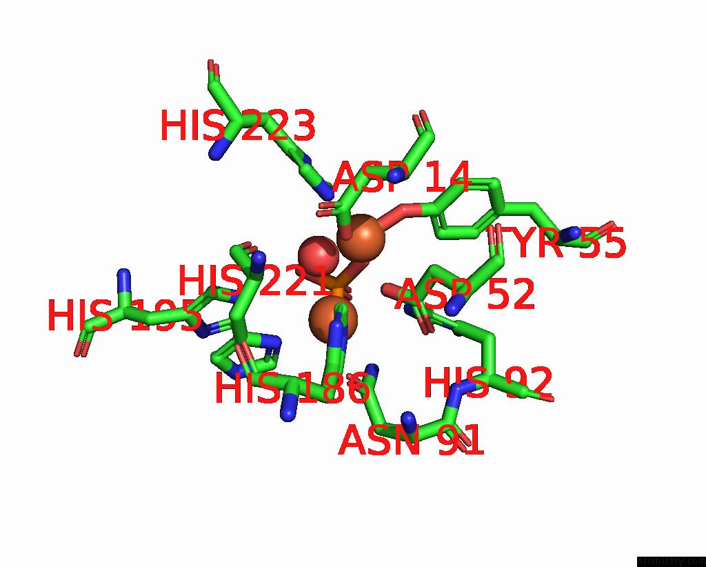
Mono view
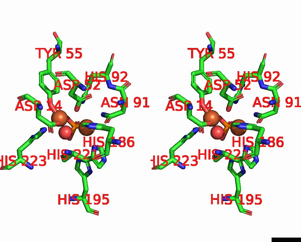
Stereo pair view
Mono view
Stereo pair view
A full contact list of Iron with other atoms in the Fe binding
site number 1 of Crystal Structure of Pig Purple Acid Phosphatase in Complex with 4-(2- Hydroxyethyl)-1-Piperazineethanesulfonic Acid (Hepes) and Glycerol within 5.0Å range:
|
Iron binding site 2 out of 2 in 7ov8
Go back to
Iron binding site 2 out
of 2 in the Crystal Structure of Pig Purple Acid Phosphatase in Complex with 4-(2- Hydroxyethyl)-1-Piperazineethanesulfonic Acid (Hepes) and Glycerol
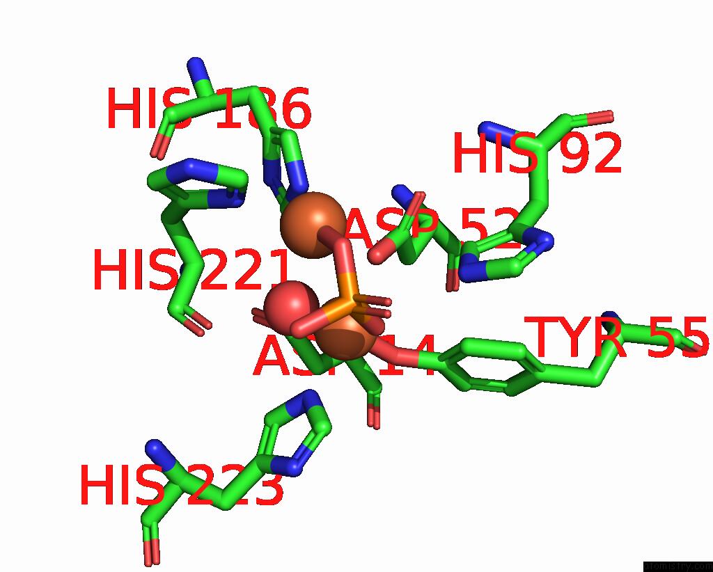
Mono view
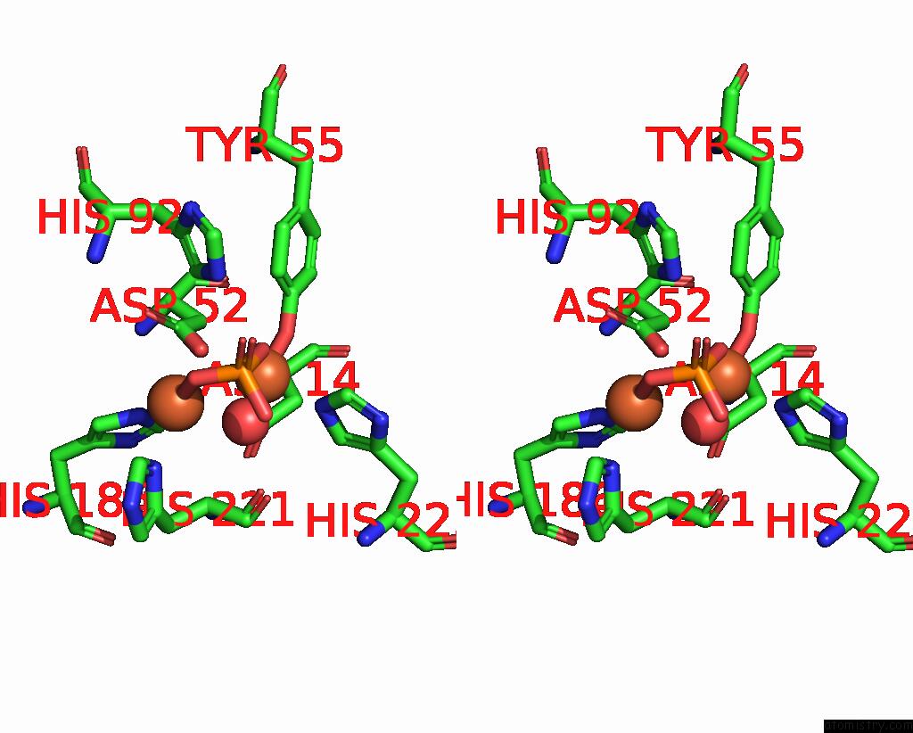
Stereo pair view
Mono view
Stereo pair view
A full contact list of Iron with other atoms in the Fe binding
site number 2 of Crystal Structure of Pig Purple Acid Phosphatase in Complex with 4-(2- Hydroxyethyl)-1-Piperazineethanesulfonic Acid (Hepes) and Glycerol within 5.0Å range:
|
Reference:
D.Feder,
S.H.Mohd-Pahmi,
W.M.Hussein,
L.W.Guddat,
R.P.Mcgeary,
G.Schenk.
Rational Design of Potent Inhibitors of A Metallohydrolase Using A Fragment-Based Approach. Chemmedchem V. 16 3342 2021.
ISSN: ESSN 1860-7187
PubMed: 34331400
DOI: 10.1002/CMDC.202100486
Page generated: Thu Aug 8 14:49:00 2024
ISSN: ESSN 1860-7187
PubMed: 34331400
DOI: 10.1002/CMDC.202100486
Last articles
Fe in 2YXOFe in 2YRS
Fe in 2YXC
Fe in 2YNM
Fe in 2YVJ
Fe in 2YP1
Fe in 2YU2
Fe in 2YU1
Fe in 2YQB
Fe in 2YOO