Iron »
PDB 7oqy-7p7j »
7oy2 »
Iron in PDB 7oy2: High Resolution Structure of Cytochrome Bd-II Oxidase From E. Coli
Enzymatic activity of High Resolution Structure of Cytochrome Bd-II Oxidase From E. Coli
All present enzymatic activity of High Resolution Structure of Cytochrome Bd-II Oxidase From E. Coli:
1.10.3.10; 7.1.1.3;
1.10.3.10; 7.1.1.3;
Iron Binding Sites:
The binding sites of Iron atom in the High Resolution Structure of Cytochrome Bd-II Oxidase From E. Coli
(pdb code 7oy2). This binding sites where shown within
5.0 Angstroms radius around Iron atom.
In total 3 binding sites of Iron where determined in the High Resolution Structure of Cytochrome Bd-II Oxidase From E. Coli, PDB code: 7oy2:
Jump to Iron binding site number: 1; 2; 3;
In total 3 binding sites of Iron where determined in the High Resolution Structure of Cytochrome Bd-II Oxidase From E. Coli, PDB code: 7oy2:
Jump to Iron binding site number: 1; 2; 3;
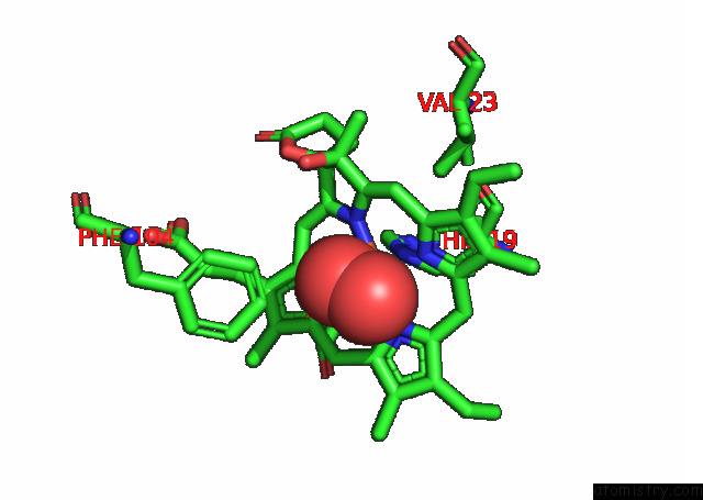
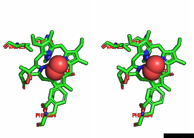
Iron binding site 1 out of 3 in 7oy2
Go back to
Iron binding site 1 out
of 3 in the High Resolution Structure of Cytochrome Bd-II Oxidase From E. Coli
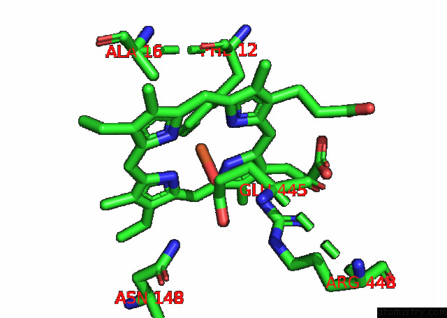
Mono view
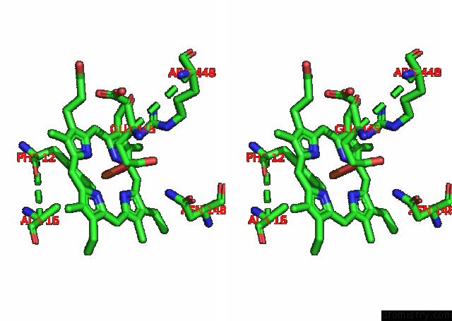
Stereo pair view
Mono view
Stereo pair view
A full contact list of Iron with other atoms in the Fe binding
site number 1 of High Resolution Structure of Cytochrome Bd-II Oxidase From E. Coli within 5.0Å range:
|
Iron binding site 2 out of 3 in 7oy2
Go back to
Iron binding site 2 out
of 3 in the High Resolution Structure of Cytochrome Bd-II Oxidase From E. Coli
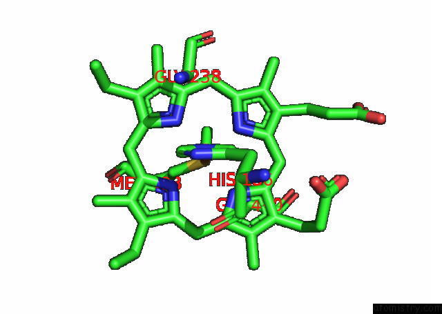
Mono view
Stereo pair view
Mono view
Stereo pair view
A full contact list of Iron with other atoms in the Fe binding
site number 2 of High Resolution Structure of Cytochrome Bd-II Oxidase From E. Coli within 5.0Å range:
|
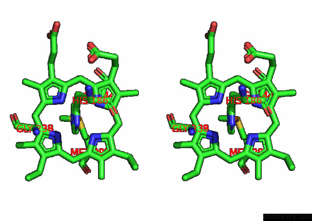
Iron binding site 3 out of 3 in 7oy2
Go back to
Iron binding site 3 out
of 3 in the High Resolution Structure of Cytochrome Bd-II Oxidase From E. Coli
Mono view
Stereo pair view
Mono view
Stereo pair view
A full contact list of Iron with other atoms in the Fe binding
site number 3 of High Resolution Structure of Cytochrome Bd-II Oxidase From E. Coli within 5.0Å range:
|
Reference:
T.N.Grund,
M.Radloff,
D.Wu,
H.G.Goojani,
L.F.Witte,
W.Josting,
S.Buschmann,
H.Muller,
I.Elamri,
S.Welsch,
H.Schwalbe,
H.Michel,
D.Bald,
S.Safarian.
Mechanistic and Structural Diversity Between Cytochrome Bd Isoforms of Escherichia Coli . Proc.Natl.Acad.Sci.Usa V. 118 2021.
ISSN: ESSN 1091-6490
PubMed: 34873041
DOI: 10.1073/PNAS.2114013118
Page generated: Thu Aug 8 14:51:01 2024
ISSN: ESSN 1091-6490
PubMed: 34873041
DOI: 10.1073/PNAS.2114013118
Last articles
Zn in 9MJ5Zn in 9HNW
Zn in 9G0L
Zn in 9FNE
Zn in 9DZN
Zn in 9E0I
Zn in 9D32
Zn in 9DAK
Zn in 8ZXC
Zn in 8ZUF