Iron »
PDB 7p7l-7pr2 »
7pq1 »
Iron in PDB 7pq1: Ligand-Free Crystal Structure of A Staphylococcal Orthologue of CYP134A1
Enzymatic activity of Ligand-Free Crystal Structure of A Staphylococcal Orthologue of CYP134A1
All present enzymatic activity of Ligand-Free Crystal Structure of A Staphylococcal Orthologue of CYP134A1:
1.14.15.13;
1.14.15.13;
Protein crystallography data
The structure of Ligand-Free Crystal Structure of A Staphylococcal Orthologue of CYP134A1, PDB code: 7pq1
was solved by
M.Snee,
C.Levy,
D.Leys,
M.Katariya,
A.W.Munro,
with X-Ray Crystallography technique. A brief refinement statistics is given in the table below:
| Resolution Low / High (Å) | 55.52 / 2.46 |
| Space group | C 2 2 21 |
| Cell size a, b, c (Å), α, β, γ (°) | 81.414, 109.222, 105.59, 90, 90, 90 |
| R / Rfree (%) | 21.4 / 25.2 |
Iron Binding Sites:
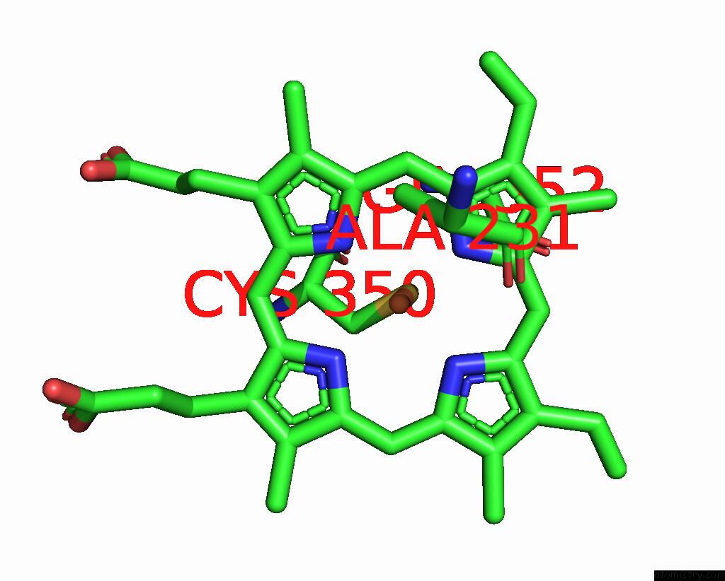
The binding sites of Iron atom in the Ligand-Free Crystal Structure of A Staphylococcal Orthologue of CYP134A1
(pdb code 7pq1). This binding sites where shown within
5.0 Angstroms radius around Iron atom.
In total only one binding site of Iron was determined in the Ligand-Free Crystal Structure of A Staphylococcal Orthologue of CYP134A1, PDB code: 7pq1:
In total only one binding site of Iron was determined in the Ligand-Free Crystal Structure of A Staphylococcal Orthologue of CYP134A1, PDB code: 7pq1:
Iron binding site 1 out of 1 in 7pq1
Go back to
Iron binding site 1 out
of 1 in the Ligand-Free Crystal Structure of A Staphylococcal Orthologue of CYP134A1
Mono view

Stereo pair view
Mono view
Stereo pair view
A full contact list of Iron with other atoms in the Fe binding
site number 1 of Ligand-Free Crystal Structure of A Staphylococcal Orthologue of CYP134A1 within 5.0Å range:
|
Reference:
M.Snee,
M.Katariya.
Crystal Structure of A Staphylococcal Orthologue of CYP134A1 (Cypx) in Complex with Cyclo-L-Leucyl-L-Leucine To Be Published.
Page generated: Thu Aug 7 02:56:06 2025
Last articles
Fe in 7S9ZFe in 7S9Y
Fe in 7S7H
Fe in 7S6T
Fe in 7S6S
Fe in 7S5C
Fe in 7S5K
Fe in 7S6R
Fe in 7S6Q
Fe in 7S5O