Iron in PDB 7pwe: Crystal Structure of the Glutaredoxin/Ferredoxin Disulfide Reductase Fusion Protein From Desulfotalea Psychrophila LSV54
Enzymatic activity of Crystal Structure of the Glutaredoxin/Ferredoxin Disulfide Reductase Fusion Protein From Desulfotalea Psychrophila LSV54
All present enzymatic activity of Crystal Structure of the Glutaredoxin/Ferredoxin Disulfide Reductase Fusion Protein From Desulfotalea Psychrophila LSV54:
1.8.7.2;
Protein crystallography data
The structure of Crystal Structure of the Glutaredoxin/Ferredoxin Disulfide Reductase Fusion Protein From Desulfotalea Psychrophila LSV54, PDB code: 7pwe
was solved by
C.Didierjean,
S.Mathiot,
N.Rouhier,
with X-Ray Crystallography technique. A brief refinement statistics is given in the table below:
Resolution Low / High (Å)
|
41.42 /
2.08
|
Space group
|
C 1 2 1
|
Cell size a, b, c (Å), α, β, γ (°)
|
123.6,
49.3,
86,
90,
116.5,
90
|
R / Rfree (%)
|
19.1 /
22.3
|
Iron Binding Sites:
The binding sites of Iron atom in the Crystal Structure of the Glutaredoxin/Ferredoxin Disulfide Reductase Fusion Protein From Desulfotalea Psychrophila LSV54
(pdb code 7pwe). This binding sites where shown within
5.0 Angstroms radius around Iron atom.
In total 8 binding sites of Iron where determined in the
Crystal Structure of the Glutaredoxin/Ferredoxin Disulfide Reductase Fusion Protein From Desulfotalea Psychrophila LSV54, PDB code: 7pwe:
Jump to Iron binding site number:
1;
2;
3;
4;
5;
6;
7;
8;
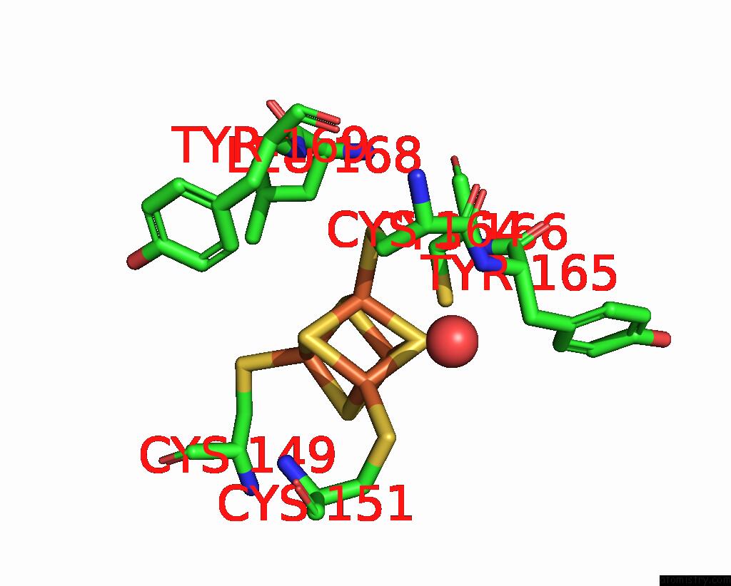
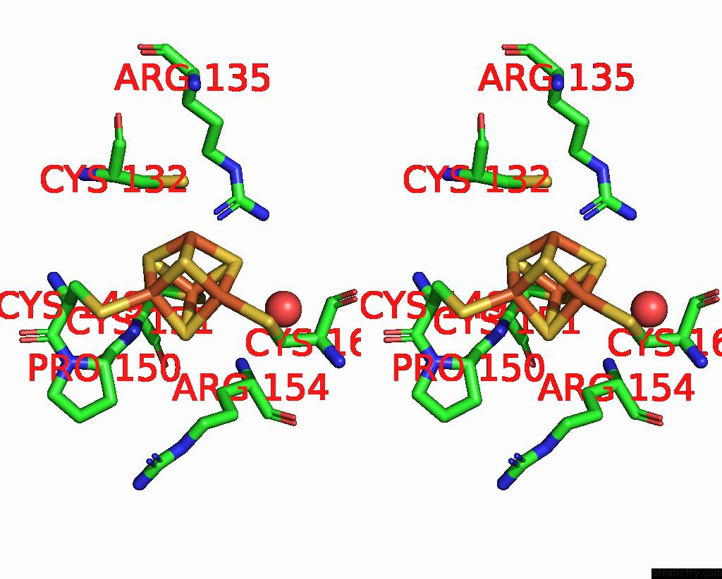
Iron binding site 1 out
of 8 in 7pwe
Go back to
Iron Binding Sites List in 7pwe
Iron binding site 1 out
of 8 in the Crystal Structure of the Glutaredoxin/Ferredoxin Disulfide Reductase Fusion Protein From Desulfotalea Psychrophila LSV54
 Mono view
 Stereo pair view
|
A full contact list of Iron with other atoms in the Fe binding
site number 1 of Crystal Structure of the Glutaredoxin/Ferredoxin Disulfide Reductase Fusion Protein From Desulfotalea Psychrophila LSV54 within 5.0Å range:
probe
|
atom
|
residue
|
distance (Å)
|
B
|
Occ
|
A:Fe201
b:21.1
occ:1.00
|
FE1
|
A:SF4201
|
0.0
|
21.1
|
1.0
|
S3
|
A:SF4201
|
2.3
|
21.6
|
1.0
|
S2
|
A:SF4201
|
2.3
|
21.2
|
1.0
|
S4
|
A:SF4201
|
2.3
|
22.0
|
1.0
|
SG
|
A:CYS164
|
2.3
|
21.6
|
1.0
|
FE2
|
A:SF4201
|
2.7
|
21.0
|
1.0
|
FE4
|
A:SF4201
|
2.7
|
20.8
|
1.0
|
FE3
|
A:SF4201
|
2.8
|
21.4
|
1.0
|
CB
|
A:CYS164
|
3.2
|
21.1
|
1.0
|
S1
|
A:SF4201
|
3.9
|
22.2
|
1.0
|
N
|
A:CYS166
|
4.1
|
22.4
|
1.0
|
CB
|
A:CYS166
|
4.2
|
24.3
|
1.0
|
N
|
A:TYR165
|
4.4
|
22.4
|
1.0
|
CB
|
A:LEU168
|
4.4
|
23.1
|
1.0
|
SG
|
A:CYS166
|
4.4
|
30.6
|
1.0
|
CA
|
A:CYS164
|
4.4
|
21.4
|
1.0
|
CD2
|
A:TYR169
|
4.5
|
25.2
|
1.0
|
C
|
A:CYS164
|
4.6
|
22.0
|
1.0
|
O
|
A:HOH347
|
4.6
|
23.6
|
1.0
|
CA
|
A:CYS166
|
4.7
|
23.2
|
1.0
|
SG
|
A:CYS151
|
4.8
|
24.1
|
1.0
|
SG
|
A:CYS149
|
4.8
|
23.2
|
1.0
|
N
|
A:LEU168
|
4.9
|
22.9
|
1.0
|
|
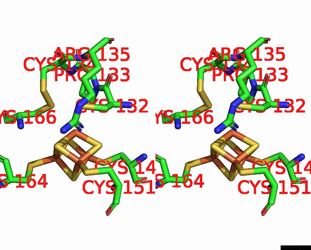
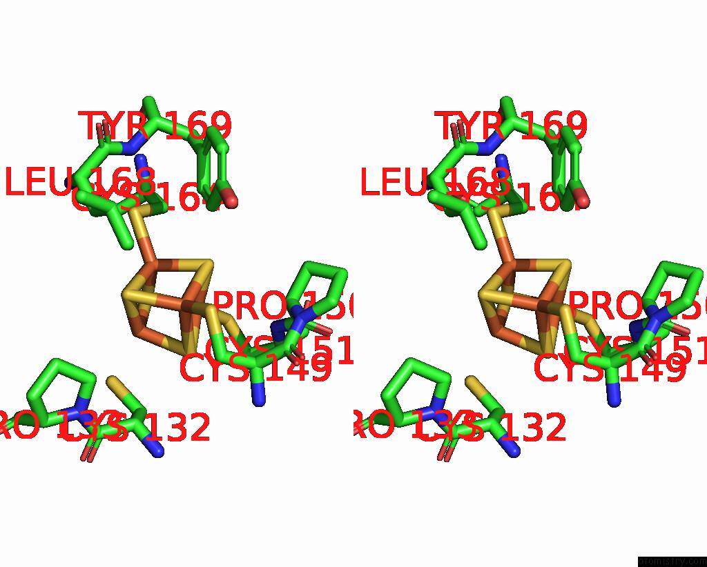
Iron binding site 2 out
of 8 in 7pwe
Go back to
Iron Binding Sites List in 7pwe
Iron binding site 2 out
of 8 in the Crystal Structure of the Glutaredoxin/Ferredoxin Disulfide Reductase Fusion Protein From Desulfotalea Psychrophila LSV54
 Mono view
 Stereo pair view
|
A full contact list of Iron with other atoms in the Fe binding
site number 2 of Crystal Structure of the Glutaredoxin/Ferredoxin Disulfide Reductase Fusion Protein From Desulfotalea Psychrophila LSV54 within 5.0Å range:
probe
|
atom
|
residue
|
distance (Å)
|
B
|
Occ
|
A:Fe201
b:21.0
occ:1.00
|
FE2
|
A:SF4201
|
0.0
|
21.0
|
1.0
|
S4
|
A:SF4201
|
2.2
|
22.0
|
1.0
|
S1
|
A:SF4201
|
2.3
|
22.2
|
1.0
|
SG
|
A:CYS151
|
2.3
|
24.1
|
1.0
|
S3
|
A:SF4201
|
2.3
|
21.6
|
1.0
|
FE1
|
A:SF4201
|
2.7
|
21.1
|
1.0
|
FE3
|
A:SF4201
|
2.7
|
21.4
|
1.0
|
FE4
|
A:SF4201
|
2.8
|
20.8
|
1.0
|
CB
|
A:CYS151
|
3.2
|
23.9
|
1.0
|
N
|
A:CYS151
|
3.5
|
21.3
|
1.0
|
S2
|
A:SF4201
|
3.8
|
21.2
|
1.0
|
NH1
|
A:ARG135
|
3.9
|
22.7
|
1.0
|
CA
|
A:CYS151
|
4.0
|
21.9
|
1.0
|
O
|
A:HOH347
|
4.0
|
23.6
|
1.0
|
SG
|
A:CYS132
|
4.6
|
26.3
|
1.0
|
CB
|
A:ARG154
|
4.6
|
22.1
|
1.0
|
CB
|
A:CYS164
|
4.6
|
21.1
|
1.0
|
C
|
A:PRO150
|
4.7
|
22.1
|
1.0
|
SG
|
A:CYS164
|
4.7
|
21.6
|
1.0
|
SG
|
A:CYS149
|
4.8
|
23.2
|
1.0
|
CA
|
A:PRO150
|
4.8
|
22.5
|
1.0
|
N
|
A:ARG154
|
4.8
|
20.5
|
1.0
|
CB
|
A:CYS132
|
4.9
|
25.8
|
1.0
|
C
|
A:CYS151
|
4.9
|
23.0
|
1.0
|
CA
|
A:ARG154
|
5.0
|
20.8
|
1.0
|
|
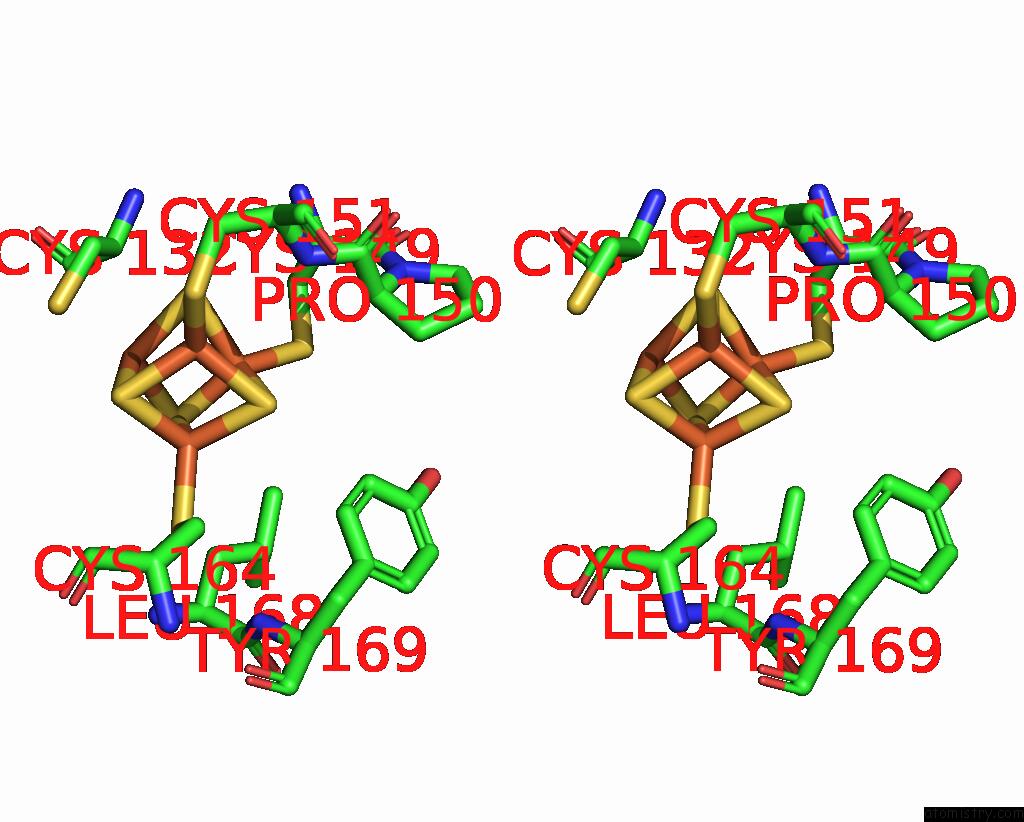
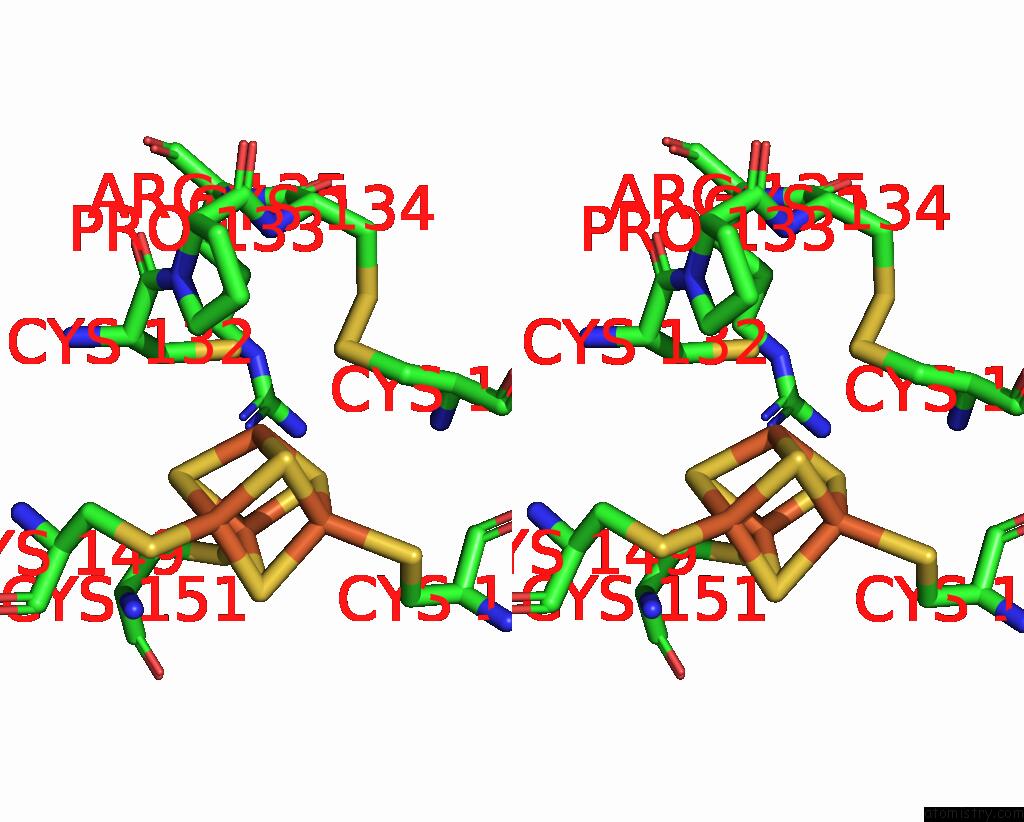
Iron binding site 3 out
of 8 in 7pwe
Go back to
Iron Binding Sites List in 7pwe
Iron binding site 3 out
of 8 in the Crystal Structure of the Glutaredoxin/Ferredoxin Disulfide Reductase Fusion Protein From Desulfotalea Psychrophila LSV54
 Mono view
 Stereo pair view
|
A full contact list of Iron with other atoms in the Fe binding
site number 3 of Crystal Structure of the Glutaredoxin/Ferredoxin Disulfide Reductase Fusion Protein From Desulfotalea Psychrophila LSV54 within 5.0Å range:
probe
|
atom
|
residue
|
distance (Å)
|
B
|
Occ
|
A:Fe201
b:21.4
occ:1.00
|
FE3
|
A:SF4201
|
0.0
|
21.4
|
1.0
|
S1
|
A:SF4201
|
2.3
|
22.2
|
1.0
|
S2
|
A:SF4201
|
2.3
|
21.2
|
1.0
|
S4
|
A:SF4201
|
2.3
|
22.0
|
1.0
|
SG
|
A:CYS132
|
2.5
|
26.3
|
1.0
|
FE2
|
A:SF4201
|
2.7
|
21.0
|
1.0
|
FE1
|
A:SF4201
|
2.8
|
21.1
|
1.0
|
FE4
|
A:SF4201
|
2.8
|
20.8
|
1.0
|
SG
|
A:CYS166
|
3.1
|
30.6
|
1.0
|
CB
|
A:CYS132
|
3.3
|
25.8
|
1.0
|
CA
|
A:CYS132
|
3.9
|
25.5
|
1.0
|
S3
|
A:SF4201
|
3.9
|
21.6
|
1.0
|
NH1
|
A:ARG135
|
4.0
|
22.7
|
1.0
|
CB
|
A:CYS166
|
4.1
|
24.3
|
1.0
|
CD
|
A:PRO133
|
4.2
|
24.0
|
1.0
|
SG
|
A:CYS134
|
4.4
|
31.6
|
1.0
|
N
|
A:CYS166
|
4.6
|
22.4
|
1.0
|
SG
|
A:CYS151
|
4.6
|
24.1
|
1.0
|
C
|
A:CYS132
|
4.6
|
25.2
|
1.0
|
N
|
A:PRO133
|
4.7
|
24.9
|
1.0
|
SG
|
A:CYS149
|
4.8
|
23.2
|
1.0
|
SG
|
A:CYS164
|
4.8
|
21.6
|
1.0
|
CA
|
A:CYS166
|
5.0
|
23.2
|
1.0
|
|
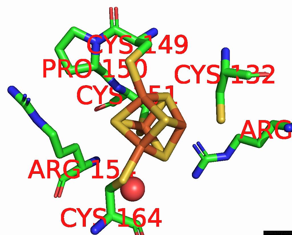
Iron binding site 4 out
of 8 in 7pwe
Go back to
Iron Binding Sites List in 7pwe
Iron binding site 4 out
of 8 in the Crystal Structure of the Glutaredoxin/Ferredoxin Disulfide Reductase Fusion Protein From Desulfotalea Psychrophila LSV54
 Mono view
 Stereo pair view
|
A full contact list of Iron with other atoms in the Fe binding
site number 4 of Crystal Structure of the Glutaredoxin/Ferredoxin Disulfide Reductase Fusion Protein From Desulfotalea Psychrophila LSV54 within 5.0Å range:
probe
|
atom
|
residue
|
distance (Å)
|
B
|
Occ
|
A:Fe201
b:20.8
occ:1.00
|
FE4
|
A:SF4201
|
0.0
|
20.8
|
1.0
|
S3
|
A:SF4201
|
2.3
|
21.6
|
1.0
|
SG
|
A:CYS149
|
2.3
|
23.2
|
1.0
|
S2
|
A:SF4201
|
2.3
|
21.2
|
1.0
|
S1
|
A:SF4201
|
2.3
|
22.2
|
1.0
|
FE1
|
A:SF4201
|
2.7
|
21.1
|
1.0
|
FE2
|
A:SF4201
|
2.8
|
21.0
|
1.0
|
FE3
|
A:SF4201
|
2.8
|
21.4
|
1.0
|
CB
|
A:CYS149
|
3.3
|
23.8
|
1.0
|
CA
|
A:CYS149
|
3.9
|
23.3
|
1.0
|
S4
|
A:SF4201
|
3.9
|
22.0
|
1.0
|
CA
|
A:PRO150
|
4.2
|
22.5
|
1.0
|
N
|
A:CYS151
|
4.4
|
21.3
|
1.0
|
CE2
|
A:TYR169
|
4.6
|
26.2
|
1.0
|
CD2
|
A:LEU168
|
4.6
|
27.4
|
1.0
|
CD2
|
A:TYR169
|
4.7
|
25.2
|
1.0
|
C
|
A:CYS149
|
4.8
|
24.0
|
1.0
|
SG
|
A:CYS164
|
4.8
|
21.6
|
1.0
|
N
|
A:PRO150
|
4.9
|
23.3
|
1.0
|
SG
|
A:CYS151
|
4.9
|
24.1
|
1.0
|
C
|
A:PRO150
|
4.9
|
22.1
|
1.0
|
N
|
A:CYS149
|
4.9
|
21.7
|
1.0
|
SG
|
A:CYS132
|
4.9
|
26.3
|
1.0
|
CB
|
A:CYS132
|
5.0
|
25.8
|
1.0
|
|
Iron binding site 5 out
of 8 in 7pwe
Go back to
Iron Binding Sites List in 7pwe
Iron binding site 5 out
of 8 in the Crystal Structure of the Glutaredoxin/Ferredoxin Disulfide Reductase Fusion Protein From Desulfotalea Psychrophila LSV54
 Mono view
 Stereo pair view
|
A full contact list of Iron with other atoms in the Fe binding
site number 5 of Crystal Structure of the Glutaredoxin/Ferredoxin Disulfide Reductase Fusion Protein From Desulfotalea Psychrophila LSV54 within 5.0Å range:
probe
|
atom
|
residue
|
distance (Å)
|
B
|
Occ
|
B:Fe200
b:23.4
occ:1.00
|
FE1
|
B:SF4200
|
0.0
|
23.4
|
1.0
|
SG
|
B:CYS151
|
2.2
|
27.9
|
1.0
|
S4
|
B:SF4200
|
2.3
|
23.7
|
1.0
|
S2
|
B:SF4200
|
2.3
|
24.0
|
1.0
|
S3
|
B:SF4200
|
2.3
|
23.4
|
1.0
|
FE4
|
B:SF4200
|
2.7
|
22.0
|
1.0
|
FE3
|
B:SF4200
|
2.7
|
23.3
|
1.0
|
FE2
|
B:SF4200
|
2.7
|
23.3
|
1.0
|
CB
|
B:CYS151
|
3.1
|
26.9
|
1.0
|
N
|
B:CYS151
|
3.5
|
26.4
|
1.0
|
S1
|
B:SF4200
|
3.9
|
23.4
|
1.0
|
CA
|
B:CYS151
|
3.9
|
26.4
|
1.0
|
NH1
|
B:ARG135
|
4.0
|
18.0
|
1.0
|
O
|
B:HOH331
|
4.1
|
26.2
|
1.0
|
CB
|
B:CYS164
|
4.6
|
25.1
|
1.0
|
C
|
B:PRO150
|
4.6
|
27.6
|
1.0
|
CB
|
B:ARG154
|
4.6
|
24.4
|
1.0
|
SG
|
B:CYS132
|
4.6
|
23.9
|
1.0
|
SG
|
B:CYS164
|
4.7
|
24.8
|
1.0
|
SG
|
B:CYS149
|
4.7
|
27.5
|
1.0
|
CA
|
B:PRO150
|
4.8
|
26.6
|
1.0
|
C
|
B:CYS151
|
4.8
|
27.1
|
1.0
|
N
|
B:ARG154
|
4.9
|
25.3
|
1.0
|
CB
|
B:CYS132
|
5.0
|
26.5
|
1.0
|
O
|
B:CYS151
|
5.0
|
27.3
|
1.0
|
|
Iron binding site 6 out
of 8 in 7pwe
Go back to
Iron Binding Sites List in 7pwe
Iron binding site 6 out
of 8 in the Crystal Structure of the Glutaredoxin/Ferredoxin Disulfide Reductase Fusion Protein From Desulfotalea Psychrophila LSV54
 Mono view
 Stereo pair view
|
A full contact list of Iron with other atoms in the Fe binding
site number 6 of Crystal Structure of the Glutaredoxin/Ferredoxin Disulfide Reductase Fusion Protein From Desulfotalea Psychrophila LSV54 within 5.0Å range:
probe
|
atom
|
residue
|
distance (Å)
|
B
|
Occ
|
B:Fe200
b:23.3
occ:1.00
|
FE2
|
B:SF4200
|
0.0
|
23.3
|
1.0
|
S3
|
B:SF4200
|
2.3
|
23.4
|
1.0
|
SG
|
B:CYS149
|
2.3
|
27.5
|
1.0
|
S1
|
B:SF4200
|
2.3
|
23.4
|
1.0
|
S4
|
B:SF4200
|
2.3
|
23.7
|
1.0
|
FE1
|
B:SF4200
|
2.7
|
23.4
|
1.0
|
FE4
|
B:SF4200
|
2.7
|
22.0
|
1.0
|
FE3
|
B:SF4200
|
2.8
|
23.3
|
1.0
|
CB
|
B:CYS149
|
3.1
|
26.8
|
1.0
|
CA
|
B:CYS149
|
3.8
|
26.2
|
1.0
|
S2
|
B:SF4200
|
3.9
|
24.0
|
1.0
|
CA
|
B:PRO150
|
4.2
|
26.6
|
1.0
|
N
|
B:CYS151
|
4.3
|
26.4
|
1.0
|
CD2
|
B:LEU168
|
4.5
|
25.7
|
1.0
|
CE2
|
B:TYR169
|
4.6
|
24.6
|
1.0
|
C
|
B:CYS149
|
4.7
|
27.3
|
1.0
|
CD2
|
B:TYR169
|
4.8
|
24.3
|
1.0
|
N
|
B:PRO150
|
4.8
|
27.1
|
1.0
|
SG
|
B:CYS151
|
4.8
|
27.9
|
1.0
|
C
|
B:PRO150
|
4.8
|
27.6
|
1.0
|
N
|
B:CYS149
|
4.8
|
25.9
|
1.0
|
SG
|
B:CYS132
|
4.8
|
23.9
|
1.0
|
CD
|
B:PRO133
|
4.9
|
24.4
|
1.0
|
SG
|
B:CYS164
|
4.9
|
24.8
|
1.0
|
CB
|
B:CYS132
|
5.0
|
26.5
|
1.0
|
|
Iron binding site 7 out
of 8 in 7pwe
Go back to
Iron Binding Sites List in 7pwe
Iron binding site 7 out
of 8 in the Crystal Structure of the Glutaredoxin/Ferredoxin Disulfide Reductase Fusion Protein From Desulfotalea Psychrophila LSV54
 Mono view
 Stereo pair view
|
A full contact list of Iron with other atoms in the Fe binding
site number 7 of Crystal Structure of the Glutaredoxin/Ferredoxin Disulfide Reductase Fusion Protein From Desulfotalea Psychrophila LSV54 within 5.0Å range:
probe
|
atom
|
residue
|
distance (Å)
|
B
|
Occ
|
B:Fe200
b:23.3
occ:1.00
|
FE3
|
B:SF4200
|
0.0
|
23.3
|
1.0
|
S2
|
B:SF4200
|
2.3
|
24.0
|
1.0
|
S1
|
B:SF4200
|
2.3
|
23.4
|
1.0
|
S4
|
B:SF4200
|
2.3
|
23.7
|
1.0
|
SG
|
B:CYS132
|
2.4
|
23.9
|
1.0
|
FE1
|
B:SF4200
|
2.7
|
23.4
|
1.0
|
FE4
|
B:SF4200
|
2.7
|
22.0
|
1.0
|
FE2
|
B:SF4200
|
2.8
|
23.3
|
1.0
|
SG
|
B:CYS166
|
3.1
|
28.6
|
1.0
|
CB
|
B:CYS132
|
3.3
|
26.5
|
1.0
|
S3
|
B:SF4200
|
3.9
|
23.4
|
1.0
|
NH1
|
B:ARG135
|
3.9
|
18.0
|
1.0
|
CA
|
B:CYS132
|
4.0
|
27.1
|
1.0
|
CD
|
B:PRO133
|
4.1
|
24.4
|
1.0
|
CB
|
B:CYS166
|
4.1
|
25.7
|
1.0
|
SG
|
B:CYS134
|
4.5
|
29.9
|
1.0
|
N
|
B:CYS166
|
4.6
|
24.3
|
1.0
|
SG
|
B:CYS151
|
4.6
|
27.9
|
1.0
|
N
|
B:PRO133
|
4.7
|
25.6
|
1.0
|
C
|
B:CYS132
|
4.7
|
27.0
|
1.0
|
SG
|
B:CYS149
|
4.8
|
27.5
|
1.0
|
SG
|
B:CYS164
|
4.9
|
24.8
|
1.0
|
|
Iron binding site 8 out
of 8 in 7pwe
Go back to
Iron Binding Sites List in 7pwe
Iron binding site 8 out
of 8 in the Crystal Structure of the Glutaredoxin/Ferredoxin Disulfide Reductase Fusion Protein From Desulfotalea Psychrophila LSV54
 Mono view
 Stereo pair view
|
A full contact list of Iron with other atoms in the Fe binding
site number 8 of Crystal Structure of the Glutaredoxin/Ferredoxin Disulfide Reductase Fusion Protein From Desulfotalea Psychrophila LSV54 within 5.0Å range:
probe
|
atom
|
residue
|
distance (Å)
|
B
|
Occ
|
B:Fe200
b:22.0
occ:1.00
|
FE4
|
B:SF4200
|
0.0
|
22.0
|
1.0
|
S2
|
B:SF4200
|
2.3
|
24.0
|
1.0
|
S1
|
B:SF4200
|
2.3
|
23.4
|
1.0
|
S3
|
B:SF4200
|
2.3
|
23.4
|
1.0
|
SG
|
B:CYS164
|
2.3
|
24.8
|
1.0
|
FE1
|
B:SF4200
|
2.7
|
23.4
|
1.0
|
FE2
|
B:SF4200
|
2.7
|
23.3
|
1.0
|
FE3
|
B:SF4200
|
2.7
|
23.3
|
1.0
|
CB
|
B:CYS164
|
3.2
|
25.1
|
1.0
|
S4
|
B:SF4200
|
3.9
|
23.7
|
1.0
|
N
|
B:CYS166
|
4.0
|
24.3
|
1.0
|
CB
|
B:CYS166
|
4.2
|
25.7
|
1.0
|
SG
|
B:CYS166
|
4.3
|
28.6
|
1.0
|
N
|
B:TYR165
|
4.3
|
25.4
|
1.0
|
CB
|
B:LEU168
|
4.3
|
23.1
|
1.0
|
CA
|
B:CYS164
|
4.4
|
25.6
|
1.0
|
C
|
B:CYS164
|
4.6
|
26.1
|
1.0
|
CD2
|
B:TYR169
|
4.6
|
24.3
|
1.0
|
CA
|
B:CYS166
|
4.7
|
24.0
|
1.0
|
O
|
B:HOH331
|
4.7
|
26.2
|
1.0
|
SG
|
B:CYS151
|
4.7
|
27.9
|
1.0
|
N
|
B:LEU168
|
4.8
|
24.0
|
1.0
|
SG
|
B:CYS149
|
4.8
|
27.5
|
1.0
|
|
Reference:
F.Zannini,
S.Mathiot,
J.Couturier,
C.Didierjean,
N.Rouhier.
Crystal Structure of the Glutaredoxin/Ferredoxin Disulfide Reductase Fusion Protein From Desulfotalea Psychrophila LSV54 Inorganics 2022.
DOI: 10.3390/INORGANICS10020024
Page generated: Thu Aug 8 17:47:43 2024
|