Iron »
PDB 7pr3-7qhm »
7qbk »
Iron in PDB 7qbk: Crystal Structure of A Second Homolog of R2-Like Ligand-Binding Oxidase in Sulfolobus Acidocaldarius (SAR2LOXII)
Protein crystallography data
The structure of Crystal Structure of A Second Homolog of R2-Like Ligand-Binding Oxidase in Sulfolobus Acidocaldarius (SAR2LOXII), PDB code: 7qbk
was solved by
H.Lebrette,
R.Diamanti,
V.Srinivas,
M.Hogbom,
with X-Ray Crystallography technique. A brief refinement statistics is given in the table below:
| Resolution Low / High (Å) | 41.91 / 2.26 |
| Space group | P 64 |
| Cell size a, b, c (Å), α, β, γ (°) | 128.029, 128.029, 41.894, 90, 90, 120 |
| R / Rfree (%) | 20.2 / 23.9 |
Other elements in 7qbk:
The structure of Crystal Structure of A Second Homolog of R2-Like Ligand-Binding Oxidase in Sulfolobus Acidocaldarius (SAR2LOXII) also contains other interesting chemical elements:
| Manganese | (Mn) | 1 atom |
Iron Binding Sites:
The binding sites of Iron atom in the Crystal Structure of A Second Homolog of R2-Like Ligand-Binding Oxidase in Sulfolobus Acidocaldarius (SAR2LOXII)
(pdb code 7qbk). This binding sites where shown within
5.0 Angstroms radius around Iron atom.
In total only one binding site of Iron was determined in the Crystal Structure of A Second Homolog of R2-Like Ligand-Binding Oxidase in Sulfolobus Acidocaldarius (SAR2LOXII), PDB code: 7qbk:
In total only one binding site of Iron was determined in the Crystal Structure of A Second Homolog of R2-Like Ligand-Binding Oxidase in Sulfolobus Acidocaldarius (SAR2LOXII), PDB code: 7qbk:
Iron binding site 1 out of 1 in 7qbk
Go back to
Iron binding site 1 out
of 1 in the Crystal Structure of A Second Homolog of R2-Like Ligand-Binding Oxidase in Sulfolobus Acidocaldarius (SAR2LOXII)
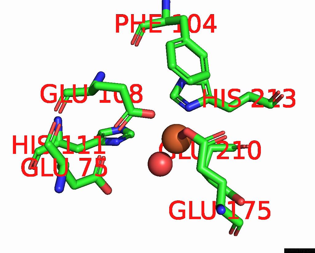
Mono view
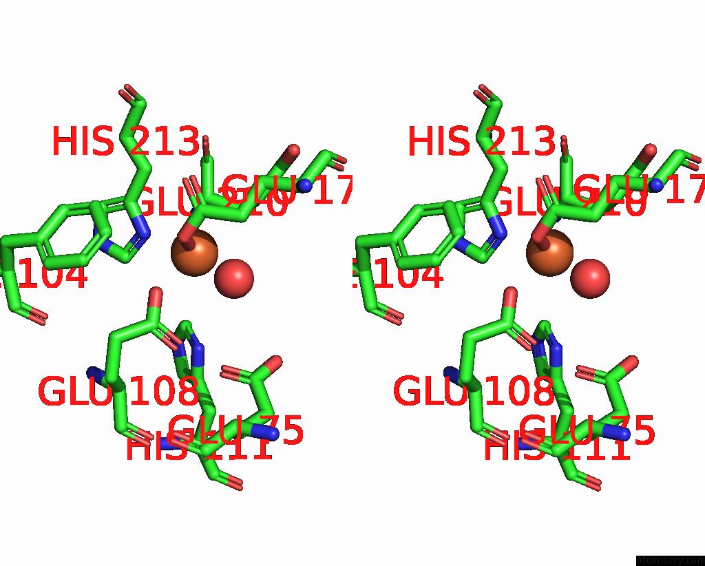
Stereo pair view
Mono view
Stereo pair view
A full contact list of Iron with other atoms in the Fe binding
site number 1 of Crystal Structure of A Second Homolog of R2-Like Ligand-Binding Oxidase in Sulfolobus Acidocaldarius (SAR2LOXII) within 5.0Å range:
|
Reference:
R.Diamanti,
V.Srinivas,
A.I.Johansson,
A.Nordstrom,
J.J.Griese,
H.Lebrette,
M.Hogbom.
Comparative Structural Analysis Provides New Insights Into the Function of R2-Like Ligand-Binding Oxidase. Febs Lett. V. 596 1600 2022.
ISSN: ISSN 0014-5793
PubMed: 35175627
DOI: 10.1002/1873-3468.14319
Page generated: Thu Aug 7 03:18:39 2025
ISSN: ISSN 0014-5793
PubMed: 35175627
DOI: 10.1002/1873-3468.14319
Last articles
K in 8HKFK in 8HKM
K in 8HIR
K in 8GYR
K in 8GR9
K in 8H5L
K in 8H5J
K in 8GPE
K in 8GNG
K in 8GPD