Iron »
PDB 7qho-7r2s »
7qke »
Iron in PDB 7qke: Crystal Structure of CYP125 From Mycobacterium Tuberculosis in Complex with Inhibitor (Surface Entropy Reduction Mutant)
Enzymatic activity of Crystal Structure of CYP125 From Mycobacterium Tuberculosis in Complex with Inhibitor (Surface Entropy Reduction Mutant)
All present enzymatic activity of Crystal Structure of CYP125 From Mycobacterium Tuberculosis in Complex with Inhibitor (Surface Entropy Reduction Mutant):
1.14.15.29;
1.14.15.29;
Protein crystallography data
The structure of Crystal Structure of CYP125 From Mycobacterium Tuberculosis in Complex with Inhibitor (Surface Entropy Reduction Mutant), PDB code: 7qke
was solved by
M.Snee,
R.Tunnicliffe,
D.Leys,
C.Levy,
M.Katariya,
with X-Ray Crystallography technique. A brief refinement statistics is given in the table below:
| Resolution Low / High (Å) | 37.70 / 2.30 |
| Space group | C 2 2 21 |
| Cell size a, b, c (Å), α, β, γ (°) | 54.01, 119.523, 145.753, 90, 90, 90 |
| R / Rfree (%) | 19.1 / 22.6 |
Iron Binding Sites:
The binding sites of Iron atom in the Crystal Structure of CYP125 From Mycobacterium Tuberculosis in Complex with Inhibitor (Surface Entropy Reduction Mutant)
(pdb code 7qke). This binding sites where shown within
5.0 Angstroms radius around Iron atom.
In total only one binding site of Iron was determined in the Crystal Structure of CYP125 From Mycobacterium Tuberculosis in Complex with Inhibitor (Surface Entropy Reduction Mutant), PDB code: 7qke:
In total only one binding site of Iron was determined in the Crystal Structure of CYP125 From Mycobacterium Tuberculosis in Complex with Inhibitor (Surface Entropy Reduction Mutant), PDB code: 7qke:
Iron binding site 1 out of 1 in 7qke
Go back to
Iron binding site 1 out
of 1 in the Crystal Structure of CYP125 From Mycobacterium Tuberculosis in Complex with Inhibitor (Surface Entropy Reduction Mutant)
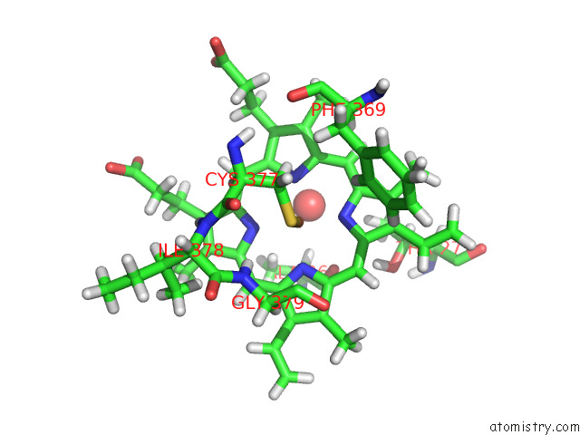
Mono view
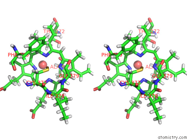
Stereo pair view
Mono view
Stereo pair view
A full contact list of Iron with other atoms in the Fe binding
site number 1 of Crystal Structure of CYP125 From Mycobacterium Tuberculosis in Complex with Inhibitor (Surface Entropy Reduction Mutant) within 5.0Å range:
|
Reference:
M.Snee,
M.Katariya.
Crystal Structure of CYP125 From Mycobacterium Tuberculosis in Complex with Inhibitor (Surface Entropy Reduction Mutant) To Be Published.
Page generated: Thu Aug 7 03:47:20 2025
Last articles
Mg in 1HN1Mg in 1HC8
Mg in 1HMV
Mg in 1HI0
Mg in 1HJK
Mg in 1HK7
Mg in 1HI8
Mg in 1HJ6
Mg in 1HBN
Mg in 1HBU