Iron »
PDB 7qho-7r2s »
7qtg »
Iron in PDB 7qtg: Crystal Structure of the Fe(II)/Alpha-Ketoglutarate Dependent Dioxygenase PLAO1
Protein crystallography data
The structure of Crystal Structure of the Fe(II)/Alpha-Ketoglutarate Dependent Dioxygenase PLAO1, PDB code: 7qtg
was solved by
P.Lukat,
M.Daum,
A.Bechthold,
O.Einsle,
with X-Ray Crystallography technique. A brief refinement statistics is given in the table below:
| Resolution Low / High (Å) | 79.74 / 2.70 |
| Space group | P 21 21 2 |
| Cell size a, b, c (Å), α, β, γ (°) | 117.923, 108.229, 65.373, 90, 90, 90 |
| R / Rfree (%) | 24 / 28.7 |
Iron Binding Sites:
The binding sites of Iron atom in the Crystal Structure of the Fe(II)/Alpha-Ketoglutarate Dependent Dioxygenase PLAO1
(pdb code 7qtg). This binding sites where shown within
5.0 Angstroms radius around Iron atom.
In total 2 binding sites of Iron where determined in the Crystal Structure of the Fe(II)/Alpha-Ketoglutarate Dependent Dioxygenase PLAO1, PDB code: 7qtg:
Jump to Iron binding site number: 1; 2;
In total 2 binding sites of Iron where determined in the Crystal Structure of the Fe(II)/Alpha-Ketoglutarate Dependent Dioxygenase PLAO1, PDB code: 7qtg:
Jump to Iron binding site number: 1; 2;
Iron binding site 1 out of 2 in 7qtg
Go back to
Iron binding site 1 out
of 2 in the Crystal Structure of the Fe(II)/Alpha-Ketoglutarate Dependent Dioxygenase PLAO1
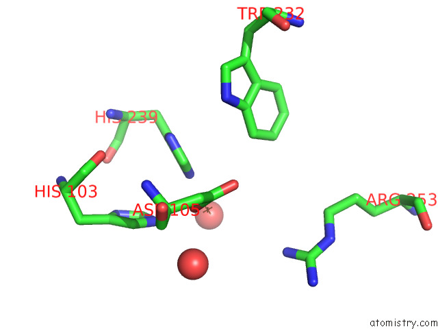
Mono view
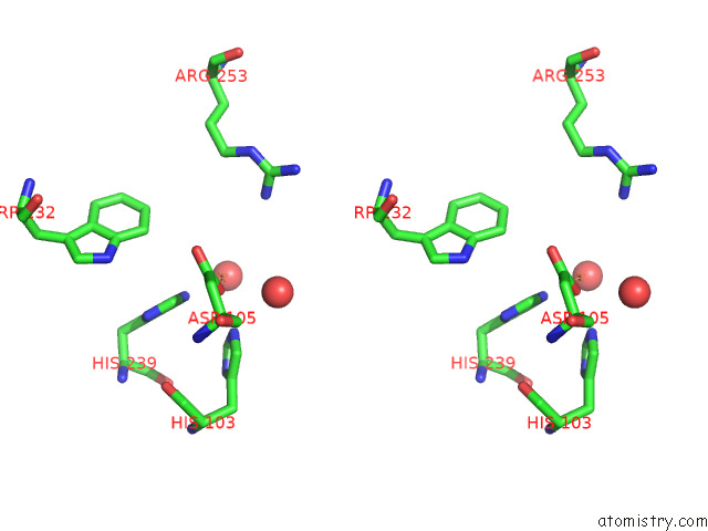
Stereo pair view
Mono view
Stereo pair view
A full contact list of Iron with other atoms in the Fe binding
site number 1 of Crystal Structure of the Fe(II)/Alpha-Ketoglutarate Dependent Dioxygenase PLAO1 within 5.0Å range:
|
Iron binding site 2 out of 2 in 7qtg
Go back to
Iron binding site 2 out
of 2 in the Crystal Structure of the Fe(II)/Alpha-Ketoglutarate Dependent Dioxygenase PLAO1
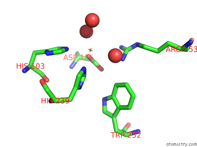
Mono view
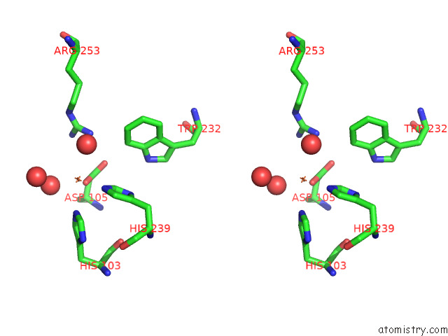
Stereo pair view
Mono view
Stereo pair view
A full contact list of Iron with other atoms in the Fe binding
site number 2 of Crystal Structure of the Fe(II)/Alpha-Ketoglutarate Dependent Dioxygenase PLAO1 within 5.0Å range:
|
Reference:
P.Lukat,
M.Daum,
D.Zechel,
A.Bechthold,
O.Einsle.
Structural Investigations on the Fe(II)/Alpha-Ketoglutarate Dependent Dioxygense PLAO1 From Streptomyces Sp. TU6071 To Be Published.
Page generated: Thu Aug 7 04:04:13 2025
Last articles
K in 9DITK in 9DJV
K in 9DIG
K in 9DID
K in 9DIC
K in 9DI8
K in 9DIB
K in 9DE5
K in 9D5W
K in 9D19