Iron »
PDB 7qho-7r2s »
7r1h »
Iron in PDB 7r1h: Crystal Structure of A Flavodiiron Protein D52K Mutant in the Reduced State From Escherichia Coli
Protein crystallography data
The structure of Crystal Structure of A Flavodiiron Protein D52K Mutant in the Reduced State From Escherichia Coli, PDB code: 7r1h
was solved by
P.T.Borges,
M.Teixeira,
C.V.Romao,
C.Frazao,
with X-Ray Crystallography technique. A brief refinement statistics is given in the table below:
| Resolution Low / High (Å) | 75.16 / 1.96 |
| Space group | I 1 2 1 |
| Cell size a, b, c (Å), α, β, γ (°) | 88.851, 64.787, 146.56, 90, 91.53, 90 |
| R / Rfree (%) | 20.1 / 24 |
Iron Binding Sites:
The binding sites of Iron atom in the Crystal Structure of A Flavodiiron Protein D52K Mutant in the Reduced State From Escherichia Coli
(pdb code 7r1h). This binding sites where shown within
5.0 Angstroms radius around Iron atom.
In total 4 binding sites of Iron where determined in the Crystal Structure of A Flavodiiron Protein D52K Mutant in the Reduced State From Escherichia Coli, PDB code: 7r1h:
Jump to Iron binding site number: 1; 2; 3; 4;
In total 4 binding sites of Iron where determined in the Crystal Structure of A Flavodiiron Protein D52K Mutant in the Reduced State From Escherichia Coli, PDB code: 7r1h:
Jump to Iron binding site number: 1; 2; 3; 4;
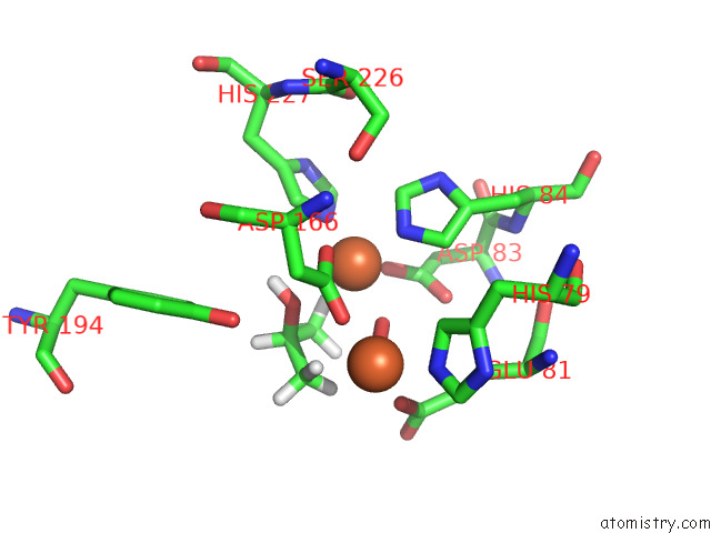
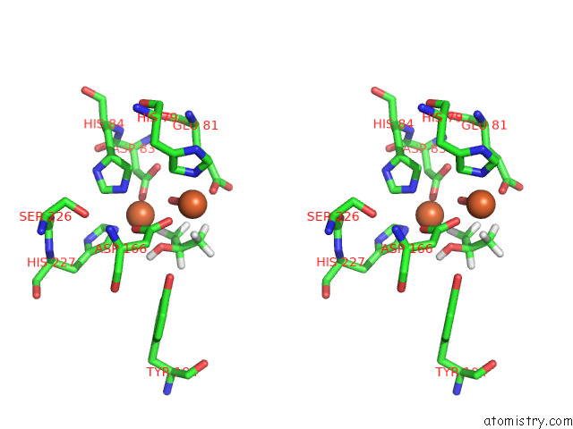
Iron binding site 1 out of 4 in 7r1h
Go back to
Iron binding site 1 out
of 4 in the Crystal Structure of A Flavodiiron Protein D52K Mutant in the Reduced State From Escherichia Coli
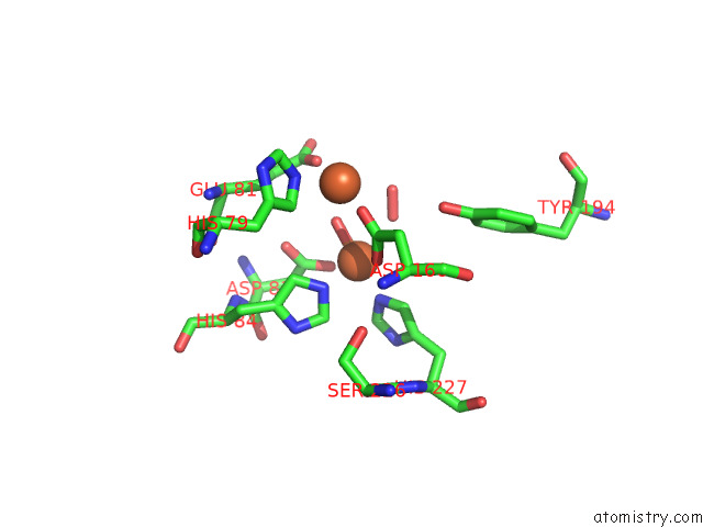
Mono view
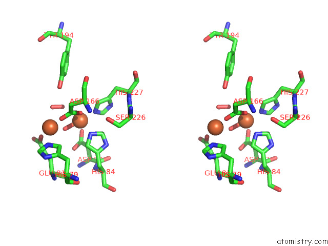
Stereo pair view
Mono view
Stereo pair view
A full contact list of Iron with other atoms in the Fe binding
site number 1 of Crystal Structure of A Flavodiiron Protein D52K Mutant in the Reduced State From Escherichia Coli within 5.0Å range:
|
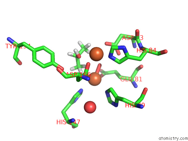
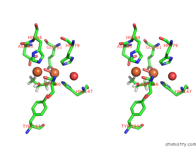
Iron binding site 2 out of 4 in 7r1h
Go back to
Iron binding site 2 out
of 4 in the Crystal Structure of A Flavodiiron Protein D52K Mutant in the Reduced State From Escherichia Coli
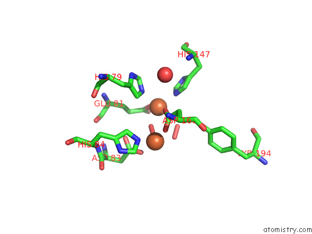
Mono view
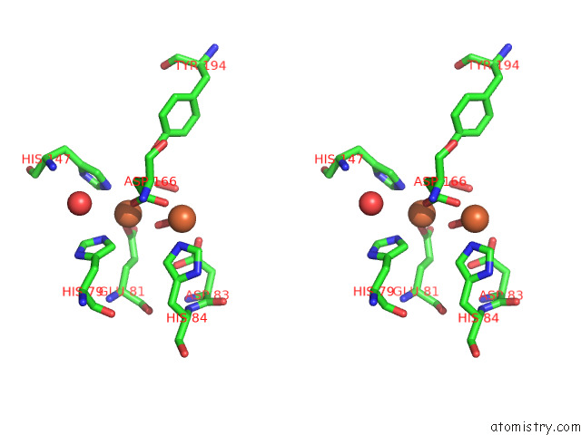
Stereo pair view
Mono view
Stereo pair view
A full contact list of Iron with other atoms in the Fe binding
site number 2 of Crystal Structure of A Flavodiiron Protein D52K Mutant in the Reduced State From Escherichia Coli within 5.0Å range:
|
Iron binding site 3 out of 4 in 7r1h
Go back to
Iron binding site 3 out
of 4 in the Crystal Structure of A Flavodiiron Protein D52K Mutant in the Reduced State From Escherichia Coli
Mono view
Stereo pair view
Mono view
Stereo pair view
A full contact list of Iron with other atoms in the Fe binding
site number 3 of Crystal Structure of A Flavodiiron Protein D52K Mutant in the Reduced State From Escherichia Coli within 5.0Å range:
|
Iron binding site 4 out of 4 in 7r1h
Go back to
Iron binding site 4 out
of 4 in the Crystal Structure of A Flavodiiron Protein D52K Mutant in the Reduced State From Escherichia Coli
Mono view
Stereo pair view
Mono view
Stereo pair view
A full contact list of Iron with other atoms in the Fe binding
site number 4 of Crystal Structure of A Flavodiiron Protein D52K Mutant in the Reduced State From Escherichia Coli within 5.0Å range:
|
Reference:
P.T.Borges,
M.Teixeira,
C.V.Romao,
C.Frazao.
Crystal Structure of A Flavodiiron Protein D52K Mutant in the Reduced State From Escherichia Coli To Be Published.
Page generated: Thu Aug 7 04:17:06 2025
Last articles
Fe in 7UT9Fe in 7UTE
Fe in 7UT8
Fe in 7UT6
Fe in 7UT7
Fe in 7USN
Fe in 7UEA
Fe in 7US8
Fe in 7US7
Fe in 7URH