Iron »
PDB 7qho-7r2s »
7r2r »
Iron in PDB 7r2r: Crystal Structure of A Flavodiiron Protein D52K/S262Y Mutant in the Reduced State From Escherichia Coli
Protein crystallography data
The structure of Crystal Structure of A Flavodiiron Protein D52K/S262Y Mutant in the Reduced State From Escherichia Coli, PDB code: 7r2r
was solved by
P.T.Borges,
M.Teixeira,
C.V.Romao,
C.Frazao,
with X-Ray Crystallography technique. A brief refinement statistics is given in the table below:
| Resolution Low / High (Å) | 44.78 / 2.20 |
| Space group | I 1 2 1 |
| Cell size a, b, c (Å), α, β, γ (°) | 88.955, 64.24, 146.85, 90, 91.38, 90 |
| R / Rfree (%) | 20.9 / 26.2 |
Iron Binding Sites:
The binding sites of Iron atom in the Crystal Structure of A Flavodiiron Protein D52K/S262Y Mutant in the Reduced State From Escherichia Coli
(pdb code 7r2r). This binding sites where shown within
5.0 Angstroms radius around Iron atom.
In total 4 binding sites of Iron where determined in the Crystal Structure of A Flavodiiron Protein D52K/S262Y Mutant in the Reduced State From Escherichia Coli, PDB code: 7r2r:
Jump to Iron binding site number: 1; 2; 3; 4;
In total 4 binding sites of Iron where determined in the Crystal Structure of A Flavodiiron Protein D52K/S262Y Mutant in the Reduced State From Escherichia Coli, PDB code: 7r2r:
Jump to Iron binding site number: 1; 2; 3; 4;
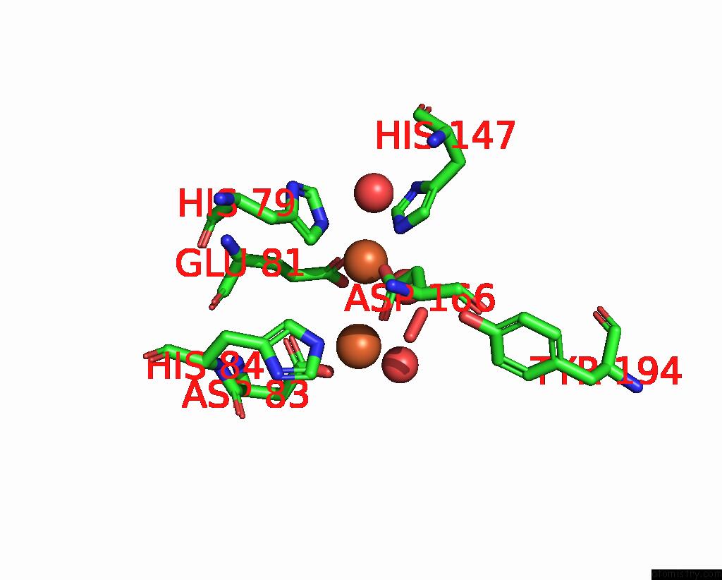
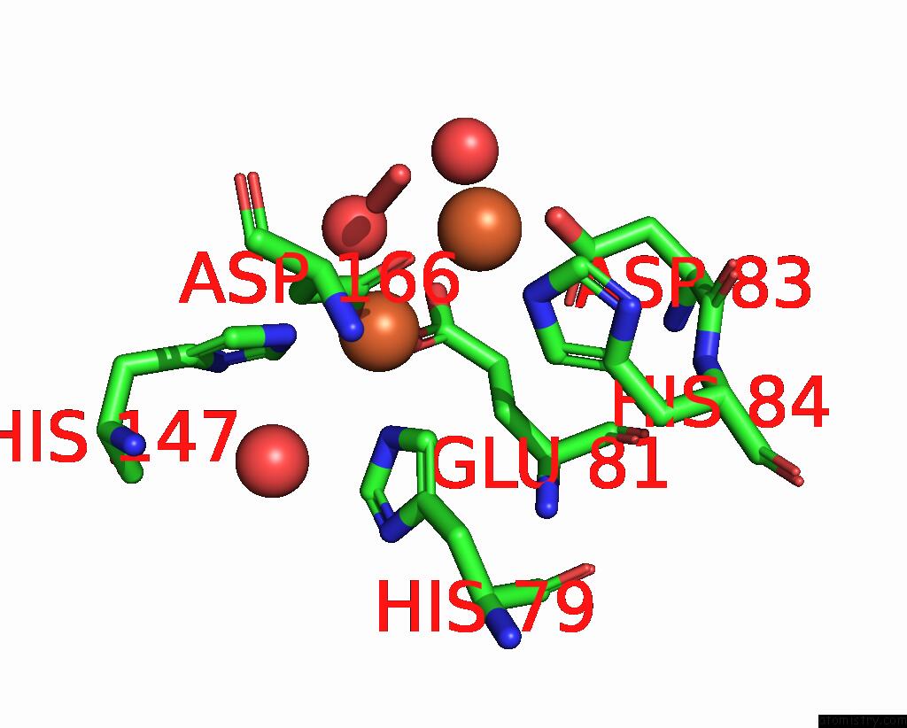
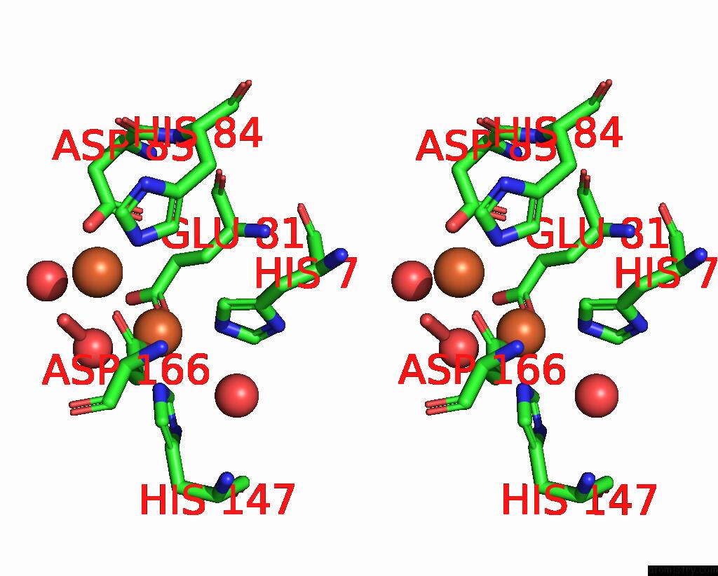
Iron binding site 1 out of 4 in 7r2r
Go back to
Iron binding site 1 out
of 4 in the Crystal Structure of A Flavodiiron Protein D52K/S262Y Mutant in the Reduced State From Escherichia Coli

Mono view

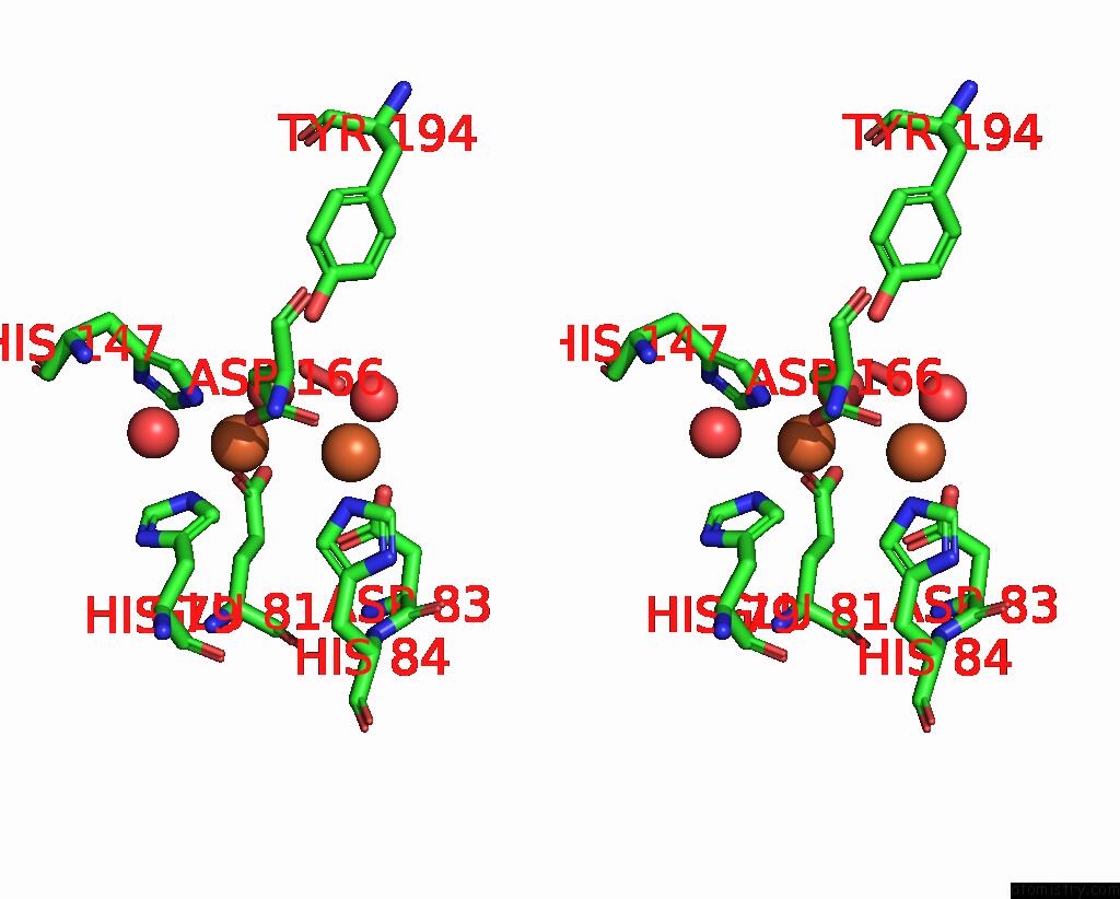
Stereo pair view
Mono view
Stereo pair view
A full contact list of Iron with other atoms in the Fe binding
site number 1 of Crystal Structure of A Flavodiiron Protein D52K/S262Y Mutant in the Reduced State From Escherichia Coli within 5.0Å range:
|
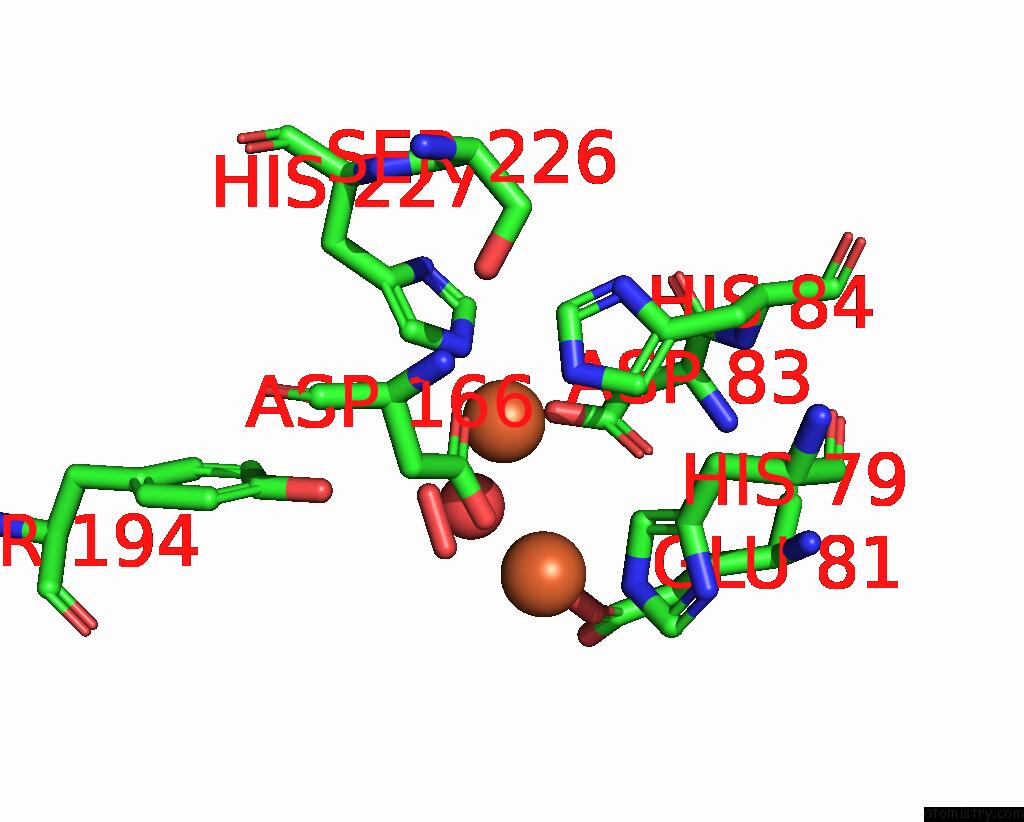
Iron binding site 2 out of 4 in 7r2r
Go back to
Iron binding site 2 out
of 4 in the Crystal Structure of A Flavodiiron Protein D52K/S262Y Mutant in the Reduced State From Escherichia Coli
Mono view
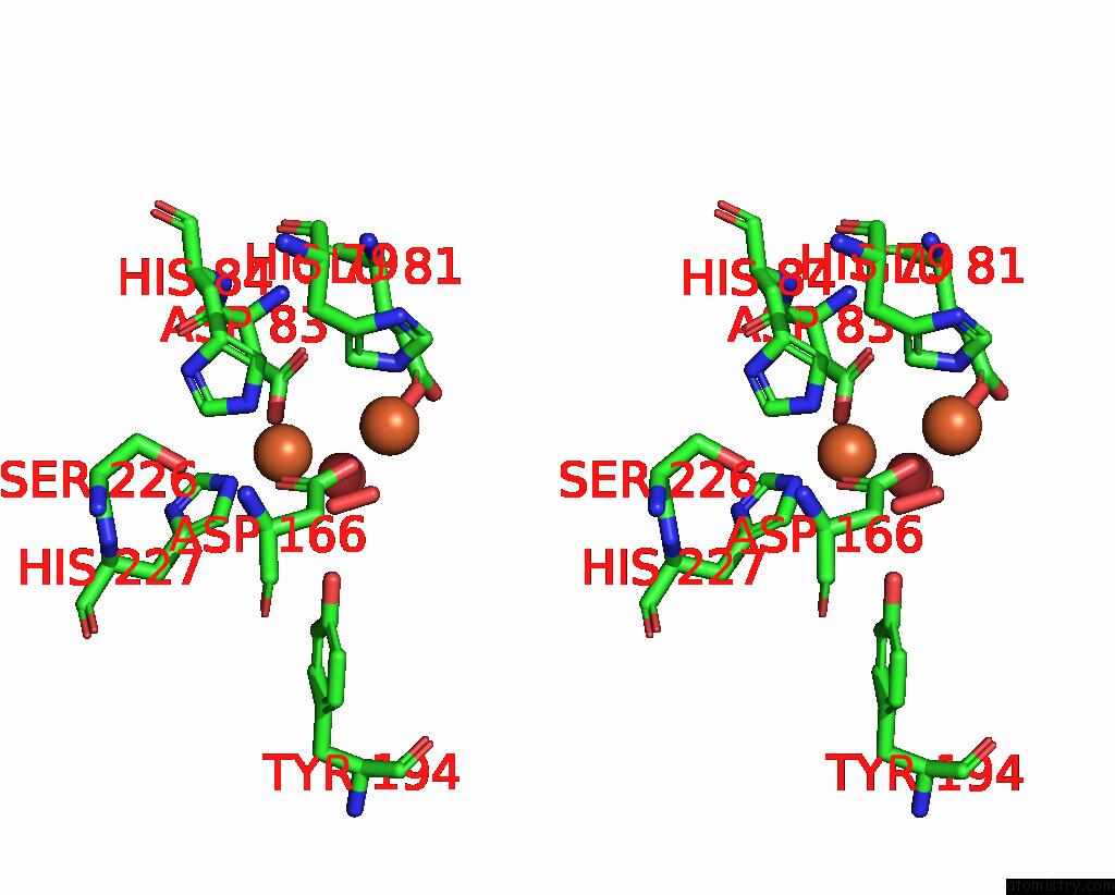
Stereo pair view
Mono view
Stereo pair view
A full contact list of Iron with other atoms in the Fe binding
site number 2 of Crystal Structure of A Flavodiiron Protein D52K/S262Y Mutant in the Reduced State From Escherichia Coli within 5.0Å range:
|
Iron binding site 3 out of 4 in 7r2r
Go back to
Iron binding site 3 out
of 4 in the Crystal Structure of A Flavodiiron Protein D52K/S262Y Mutant in the Reduced State From Escherichia Coli
Mono view
Stereo pair view
Mono view
Stereo pair view
A full contact list of Iron with other atoms in the Fe binding
site number 3 of Crystal Structure of A Flavodiiron Protein D52K/S262Y Mutant in the Reduced State From Escherichia Coli within 5.0Å range:
|
Iron binding site 4 out of 4 in 7r2r
Go back to
Iron binding site 4 out
of 4 in the Crystal Structure of A Flavodiiron Protein D52K/S262Y Mutant in the Reduced State From Escherichia Coli
Mono view
Stereo pair view
Mono view
Stereo pair view
A full contact list of Iron with other atoms in the Fe binding
site number 4 of Crystal Structure of A Flavodiiron Protein D52K/S262Y Mutant in the Reduced State From Escherichia Coli within 5.0Å range:
|
Reference:
P.T.Borges,
M.Teixeira,
C.V.Romao,
C.Frazao.
Crystal Structure of A Flavodiiron Protein D52K/S262Y Mutant in the Reduced State From Escherichia Coli To Be Published.
Page generated: Thu Aug 7 04:19:20 2025
Last articles
K in 9DITK in 9DJV
K in 9DIG
K in 9DID
K in 9DIC
K in 9DI8
K in 9DIB
K in 9DE5
K in 9D5W
K in 9D19