Atomistry » Iron » PDB 7r2r-7rkr » 7r3v
Atomistry »
  Iron »
    PDB 7r2r-7rkr »
      7r3v »

Iron in PDB 7r3v: Crystal Structure of Bovine Cytochrome BC1 in Complex with Inhibitor Ck-2-67.

Enzymatic activity of Crystal Structure of Bovine Cytochrome BC1 in Complex with Inhibitor Ck-2-67.

All present enzymatic activity of Crystal Structure of Bovine Cytochrome BC1 in Complex with Inhibitor Ck-2-67.:
7.1.1.8;

Protein crystallography data

The structure of Crystal Structure of Bovine Cytochrome BC1 in Complex with Inhibitor Ck-2-67., PDB code: 7r3v was solved by N.Pinthong, K.Amporndanai, P.M.O'neill, S.S.Hasnain, S.Antonyuk, with X-Ray Crystallography technique. A brief refinement statistics is given in the table below:

Resolution Low / High (Å) 49.86 / 3.20
Space group P 65 2 2
Cell size a, b, c (Å), α, β, γ (°) 209.589, 209.589, 342.429, 90, 90, 120
R / Rfree (%) 20.5 / 25.6

Other elements in 7r3v:

The structure of Crystal Structure of Bovine Cytochrome BC1 in Complex with Inhibitor Ck-2-67. also contains other interesting chemical elements:

Fluorine (F) 3 atoms

Iron Binding Sites:

The binding sites of Iron atom in the Crystal Structure of Bovine Cytochrome BC1 in Complex with Inhibitor Ck-2-67. (pdb code 7r3v). This binding sites where shown within 5.0 Angstroms radius around Iron atom.
In total 5 binding sites of Iron where determined in the Crystal Structure of Bovine Cytochrome BC1 in Complex with Inhibitor Ck-2-67., PDB code: 7r3v:
Jump to Iron binding site number: 1; 2; 3; 4; 5;

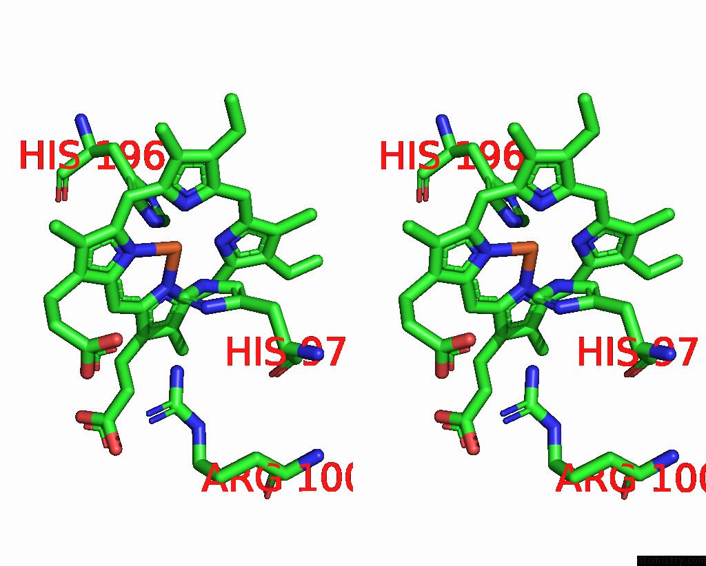
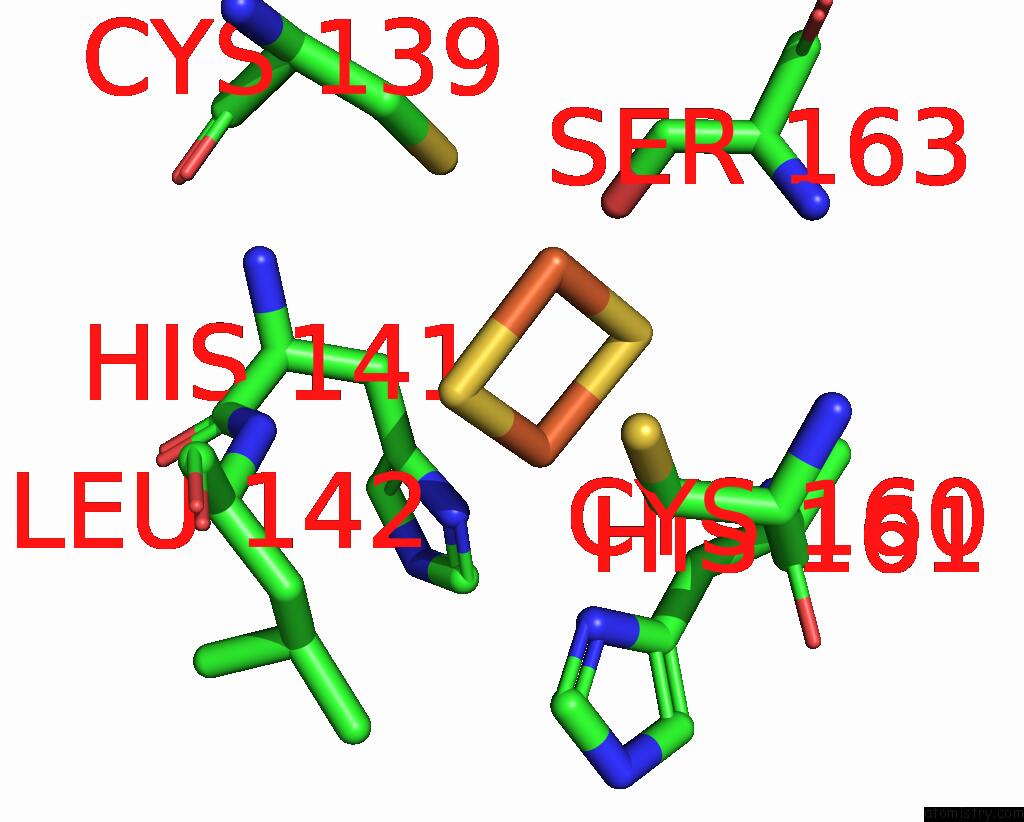
Iron binding site 1 out of 5 in 7r3v

Go back to Iron Binding Sites List in 7r3v
Iron binding site 1 out of 5 in the Crystal Structure of Bovine Cytochrome BC1 in Complex with Inhibitor Ck-2-67.


Mono view


Stereo pair view

A full contact list of Iron with other atoms in the Fe binding site number 1 of Crystal Structure of Bovine Cytochrome BC1 in Complex with Inhibitor Ck-2-67. within 5.0Å range:
probe atom residue distance (Å) B Occ
C:Fe401

b:62.4
occ:1.00
FE C:HEM401 0.0 62.4 1.0
ND C:HEM401 1.9 60.5 1.0
NA C:HEM401 2.0 70.5 1.0
NC C:HEM401 2.0 65.2 1.0
NB C:HEM401 2.1 63.9 1.0
NE2 C:HIS83 2.2 59.6 1.0
NE2 C:HIS182 2.2 50.9 1.0
C4D C:HEM401 2.9 67.6 1.0
C1D C:HEM401 2.9 57.0 1.0
C1A C:HEM401 3.0 69.8 1.0
CE1 C:HIS83 3.0 65.1 1.0
C4C C:HEM401 3.0 67.3 1.0
CE1 C:HIS182 3.0 58.0 1.0
C4B C:HEM401 3.1 69.1 1.0
C4A C:HEM401 3.1 72.3 1.0
C1B C:HEM401 3.1 70.3 1.0
C1C C:HEM401 3.1 67.8 1.0
CD2 C:HIS182 3.2 53.8 1.0
CD2 C:HIS83 3.2 60.3 1.0
CHA C:HEM401 3.3 71.6 1.0
CHD C:HEM401 3.4 62.0 1.0
CHC C:HEM401 3.5 71.2 1.0
CHB C:HEM401 3.5 71.7 1.0
ND1 C:HIS83 4.1 77.2 1.0
C2D C:HEM401 4.2 63.5 1.0
C2A C:HEM401 4.2 66.7 1.0
ND1 C:HIS182 4.2 65.1 1.0
C3D C:HEM401 4.2 69.4 1.0
C3A C:HEM401 4.2 68.6 1.0
C3C C:HEM401 4.2 69.7 1.0
C2C C:HEM401 4.3 73.4 1.0
CG C:HIS83 4.3 67.7 1.0
C2B C:HEM401 4.3 70.6 1.0
CG C:HIS182 4.3 57.2 1.0
C3B C:HEM401 4.3 72.3 1.0
NE2 C:GLN44 4.9 64.4 1.0
CA C:GLY130 4.9 70.0 1.0

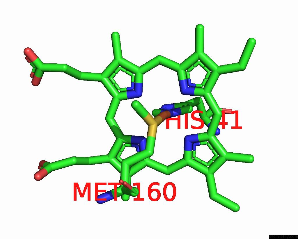
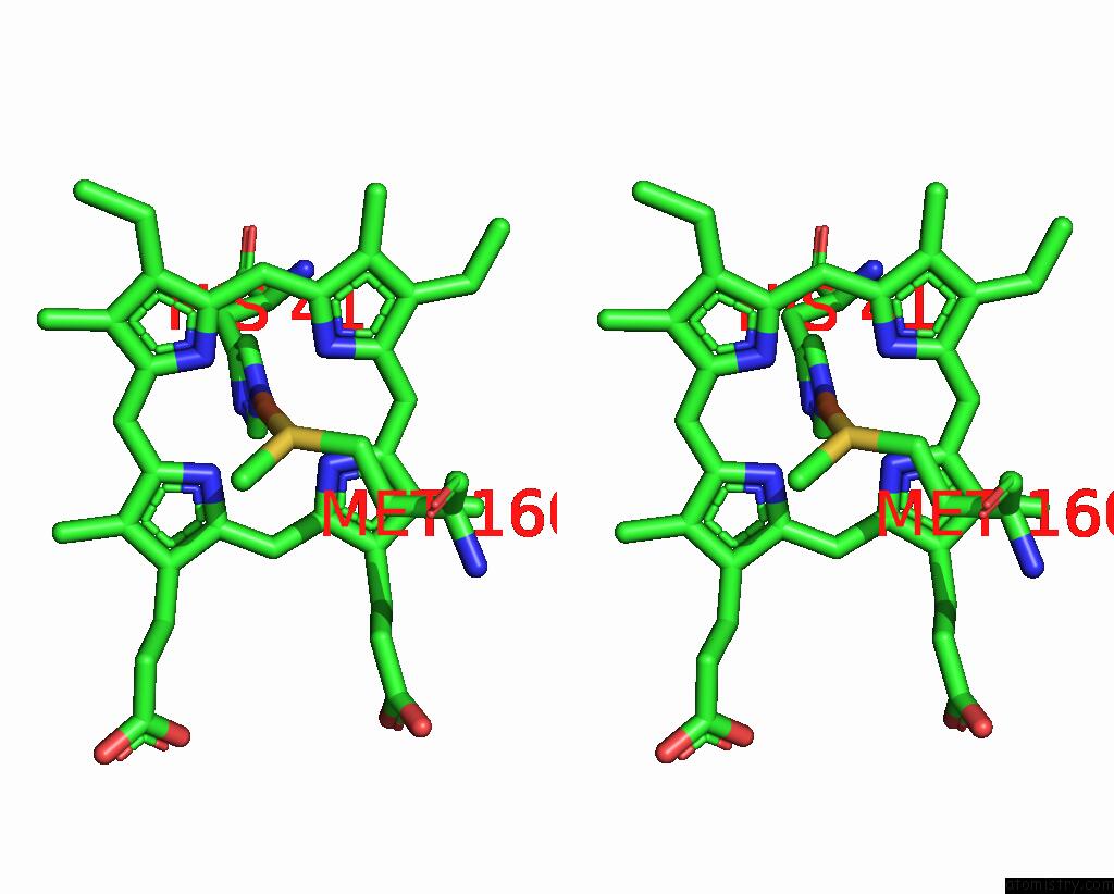
Iron binding site 2 out of 5 in 7r3v

Go back to Iron Binding Sites List in 7r3v
Iron binding site 2 out of 5 in the Crystal Structure of Bovine Cytochrome BC1 in Complex with Inhibitor Ck-2-67.


Mono view


Stereo pair view

A full contact list of Iron with other atoms in the Fe binding site number 2 of Crystal Structure of Bovine Cytochrome BC1 in Complex with Inhibitor Ck-2-67. within 5.0Å range:
probe atom residue distance (Å) B Occ
C:Fe402

b:51.8
occ:1.00
FE C:HEM402 0.0 51.8 1.0
ND C:HEM402 1.9 56.8 1.0
NA C:HEM402 2.0 64.3 1.0
NB C:HEM402 2.0 55.8 1.0
NC C:HEM402 2.0 56.3 1.0
NE2 C:HIS196 2.2 43.8 1.0
NE2 C:HIS97 2.2 55.5 1.0
C4D C:HEM402 2.9 57.1 1.0
C1D C:HEM402 2.9 56.5 1.0
C1A C:HEM402 3.0 63.9 1.0
C4C C:HEM402 3.0 56.0 1.0
C4B C:HEM402 3.0 67.3 1.0
CE1 C:HIS97 3.0 60.5 1.0
C1B C:HEM402 3.0 58.6 1.0
C1C C:HEM402 3.1 59.5 1.0
C4A C:HEM402 3.1 63.9 1.0
CD2 C:HIS196 3.1 46.1 1.0
CE1 C:HIS196 3.2 52.9 1.0
CD2 C:HIS97 3.2 52.6 1.0
CHD C:HEM402 3.3 54.1 1.0
CHA C:HEM402 3.4 56.6 1.0
CHC C:HEM402 3.4 64.2 1.0
CHB C:HEM402 3.5 66.8 1.0
ND1 C:HIS97 4.1 63.3 1.0
C3D C:HEM402 4.2 63.3 1.0
C2A C:HEM402 4.2 56.6 1.0
C2D C:HEM402 4.2 62.5 1.0
C3C C:HEM402 4.2 60.8 1.0
CG C:HIS196 4.2 51.1 1.0
C2C C:HEM402 4.2 62.3 1.0
ND1 C:HIS196 4.2 55.0 1.0
CG C:HIS97 4.3 56.7 1.0
C3A C:HEM402 4.3 54.9 1.0
C3B C:HEM402 4.3 67.7 1.0
C2B C:HEM402 4.3 59.0 1.0
NH2 C:ARG100 5.0 68.0 1.0

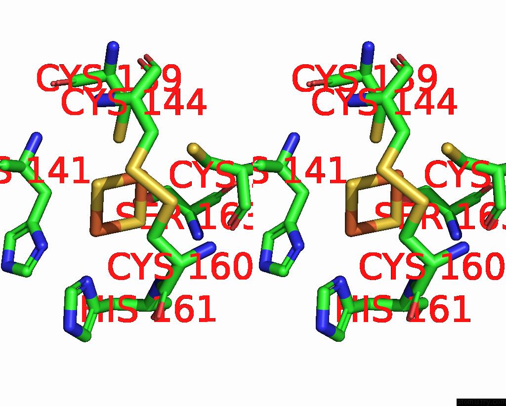
Iron binding site 3 out of 5 in 7r3v

Go back to Iron Binding Sites List in 7r3v
Iron binding site 3 out of 5 in the Crystal Structure of Bovine Cytochrome BC1 in Complex with Inhibitor Ck-2-67.


Mono view


Stereo pair view

A full contact list of Iron with other atoms in the Fe binding site number 3 of Crystal Structure of Bovine Cytochrome BC1 in Complex with Inhibitor Ck-2-67. within 5.0Å range:
probe atom residue distance (Å) B Occ
D:Fe501

b:90.7
occ:1.00
FE D:HEC501 0.0 90.7 1.0
NE2 D:HIS41 1.8 120.0 1.0
NB D:HEC501 2.1 78.6 1.0
NA D:HEC501 2.1 86.1 1.0
ND D:HEC501 2.1 84.8 1.0
NC D:HEC501 2.1 102.7 1.0
SD D:MET160 2.3 107.1 1.0
CE1 D:HIS41 2.8 119.3 1.0
CD2 D:HIS41 2.9 120.0 1.0
C4B D:HEC501 3.0 85.4 1.0
C1B D:HEC501 3.1 78.4 1.0
C4A D:HEC501 3.1 92.2 1.0
C4D D:HEC501 3.1 91.3 1.0
C1A D:HEC501 3.1 83.8 1.0
C1C D:HEC501 3.1 108.2 1.0
C1D D:HEC501 3.1 79.8 1.0
C4C D:HEC501 3.1 108.5 1.0
CHC D:HEC501 3.4 100.3 1.0
CHB D:HEC501 3.4 81.2 1.0
CHA D:HEC501 3.4 93.1 1.0
CHD D:HEC501 3.5 94.3 1.0
CE D:MET160 3.6 101.7 1.0
CG D:MET160 3.7 109.9 1.0
ND1 D:HIS41 3.9 124.4 1.0
CG D:HIS41 4.0 116.8 1.0
C3B D:HEC501 4.3 85.6 1.0
C2B D:HEC501 4.4 82.1 1.0
C3A D:HEC501 4.4 94.2 1.0
C3D D:HEC501 4.4 94.6 1.0
C2D D:HEC501 4.4 89.8 1.0
C2A D:HEC501 4.5 82.4 1.0
CB D:MET160 4.5 103.8 1.0
C3C D:HEC501 4.5 107.9 1.0
C2C D:HEC501 4.5 111.1 1.0

Iron binding site 4 out of 5 in 7r3v

Go back to Iron Binding Sites List in 7r3v
Iron binding site 4 out of 5 in the Crystal Structure of Bovine Cytochrome BC1 in Complex with Inhibitor Ck-2-67.


Mono view


Stereo pair view

A full contact list of Iron with other atoms in the Fe binding site number 4 of Crystal Structure of Bovine Cytochrome BC1 in Complex with Inhibitor Ck-2-67. within 5.0Å range:
probe atom residue distance (Å) B Occ
E:Fe201

b:144.8
occ:0.50
FE1 E:FES201 0.0 144.8 0.5
S2 E:FES201 2.2 185.9 0.5
S1 E:FES201 2.2 211.0 1.0
SG E:CYS158 2.6 144.5 0.5
SG E:CYS139 2.6 192.7 1.0
CB E:CYS158 2.9 155.0 1.0
FE2 E:FES201 3.0 205.8 1.0
CB E:CYS139 3.3 149.9 1.0
CB E:CYS144 3.9 176.2 1.0
CB E:CYS160 4.0 153.7 1.0
CB E:SER163 4.2 251.9 1.0
OG E:SER163 4.2 173.0 1.0
SG E:CYS144 4.3 168.8 1.0
SG E:CYS160 4.3 162.2 1.0
CA E:CYS158 4.3 164.9 0.5
O E:CYS158 4.4 150.8 1.0
C E:CYS158 4.5 150.9 1.0
N E:HIS161 4.6 245.8 1.0
N E:CYS160 4.6 207.5 1.0
N E:CYS144 4.7 184.4 1.0
CA E:CYS139 4.7 159.3 1.0
CB E:HIS141 4.8 172.3 1.0
CA E:CYS160 4.8 188.5 1.0
CA E:CYS144 4.9 175.8 1.0

Iron binding site 5 out of 5 in 7r3v

Go back to Iron Binding Sites List in 7r3v
Iron binding site 5 out of 5 in the Crystal Structure of Bovine Cytochrome BC1 in Complex with Inhibitor Ck-2-67.


Mono view


Stereo pair view

A full contact list of Iron with other atoms in the Fe binding site number 5 of Crystal Structure of Bovine Cytochrome BC1 in Complex with Inhibitor Ck-2-67. within 5.0Å range:
probe atom residue distance (Å) B Occ
E:Fe201

b:205.8
occ:1.00
FE2 E:FES201 0.0 205.8 1.0
S2 E:FES201 2.2 185.9 0.5
S1 E:FES201 2.2 211.0 1.0
ND1 E:HIS161 2.7 187.5 1.0
ND1 E:HIS141 2.7 194.8 1.0
FE1 E:FES201 3.0 144.8 0.5
CB E:HIS161 3.2 236.7 1.0
CG E:HIS161 3.3 211.2 1.0
CG E:HIS141 3.5 167.4 1.0
CB E:HIS141 3.5 172.3 1.0
N E:HIS161 3.5 245.8 1.0
CE1 E:HIS141 3.7 201.4 1.0
CB E:CYS160 3.8 153.7 1.0
CE1 E:HIS161 3.8 172.4 1.0
CA E:HIS161 3.9 242.6 1.0
N E:LEU142 4.1 185.6 1.0
C E:CYS160 4.2 208.0 1.0
CB E:LEU142 4.3 150.0 1.0
SG E:CYS139 4.5 192.7 1.0
CG E:LEU142 4.5 126.9 1.0
CD2 E:HIS161 4.6 198.0 1.0
CA E:CYS160 4.6 188.5 1.0
C E:HIS161 4.7 221.6 1.0
OG E:SER163 4.7 173.0 1.0
CD2 E:HIS141 4.7 171.6 1.0
CA E:HIS141 4.7 168.1 1.0
CA E:LEU142 4.8 177.9 1.0
NE2 E:HIS161 4.8 189.0 1.0
NE2 E:HIS141 4.8 201.2 1.0
C E:HIS141 4.8 164.7 1.0
CD1 E:LEU142 4.9 107.5 1.0
O E:CYS160 5.0 191.8 1.0

Reference:

K.Amporndanai, N.Pinthong, P.M.O'neill, W.D.Hong, R.K.Amewu, C.Pidathala, N.G.Berry, S.C.Leung, S.A.Ward, G.A.Biagini, S.S.Hasnain, S.V.Antonyuk. Targeting the Ubiquinol-Reduction (Q I ) Site of the Mitochondrial Cytochrome Bc 1 Complex For the Development of Next Generation Quinolone Antimalarials. Biology (Basel) V. 11 2022.
ISSN: ESSN 2079-7737
PubMed: 35892964
DOI: 10.3390/BIOLOGY11081109
Page generated: Wed Apr 5 04:49:06 2023

Last articles

Zn in 8WB0
Zn in 8WAX
Zn in 8WAU
Zn in 8WAZ
Zn in 8WAY
Zn in 8WAV
Zn in 8WAW
Zn in 8WAT
Zn in 8W7M
Zn in 8WD3
© Copyright 2008-2020 by atomistry.com
Home   |    Site Map   |    Copyright   |    Contact us   |    Privacy