Iron in PDB 7rcv: High-Resolution Structure of Photosystem II From the Mesophilic Cyanobacterium, Synechocystis Sp. Pcc 6803
Enzymatic activity of High-Resolution Structure of Photosystem II From the Mesophilic Cyanobacterium, Synechocystis Sp. Pcc 6803
All present enzymatic activity of High-Resolution Structure of Photosystem II From the Mesophilic Cyanobacterium, Synechocystis Sp. Pcc 6803:
1.10.3.9;
Other elements in 7rcv:
The structure of High-Resolution Structure of Photosystem II From the Mesophilic Cyanobacterium, Synechocystis Sp. Pcc 6803 also contains other interesting chemical elements:
Iron Binding Sites:
The binding sites of Iron atom in the High-Resolution Structure of Photosystem II From the Mesophilic Cyanobacterium, Synechocystis Sp. Pcc 6803
(pdb code 7rcv). This binding sites where shown within
5.0 Angstroms radius around Iron atom.
In total 6 binding sites of Iron where determined in the
High-Resolution Structure of Photosystem II From the Mesophilic Cyanobacterium, Synechocystis Sp. Pcc 6803, PDB code: 7rcv:
Jump to Iron binding site number:
1;
2;
3;
4;
5;
6;
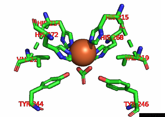
Iron binding site 1 out
of 6 in 7rcv
Go back to
Iron Binding Sites List in 7rcv
Iron binding site 1 out
of 6 in the High-Resolution Structure of Photosystem II From the Mesophilic Cyanobacterium, Synechocystis Sp. Pcc 6803
Mono view
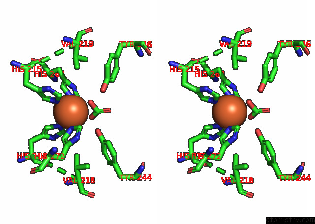
Stereo pair view
|
A full contact list of Iron with other atoms in the Fe binding
site number 1 of High-Resolution Structure of Photosystem II From the Mesophilic Cyanobacterium, Synechocystis Sp. Pcc 6803 within 5.0Å range:
probe
|
atom
|
residue
|
distance (Å)
|
B
|
Occ
|
A:Fe402
b:3.3
occ:1.00
|
NE2
|
A:HIS215
|
2.3
|
3.6
|
1.0
|
O1
|
A:BCT418
|
2.4
|
4.2
|
1.0
|
O3
|
A:BCT418
|
2.4
|
4.2
|
1.0
|
NE2
|
D:HIS268
|
2.4
|
3.7
|
1.0
|
NE2
|
A:HIS272
|
2.4
|
3.5
|
1.0
|
NE2
|
D:HIS214
|
2.4
|
10.5
|
1.0
|
C
|
A:BCT418
|
2.8
|
4.2
|
1.0
|
CD2
|
A:HIS215
|
3.2
|
3.6
|
1.0
|
CD2
|
D:HIS214
|
3.3
|
10.5
|
1.0
|
CD2
|
D:HIS268
|
3.3
|
3.7
|
1.0
|
CE1
|
A:HIS272
|
3.3
|
3.5
|
1.0
|
CE1
|
A:HIS215
|
3.3
|
3.6
|
1.0
|
CE1
|
D:HIS268
|
3.4
|
3.7
|
1.0
|
CD2
|
A:HIS272
|
3.4
|
3.5
|
1.0
|
CE1
|
D:HIS214
|
3.5
|
10.5
|
1.0
|
O2
|
A:BCT418
|
4.1
|
4.2
|
1.0
|
CG
|
A:HIS215
|
4.4
|
3.6
|
1.0
|
ND1
|
A:HIS215
|
4.4
|
3.6
|
1.0
|
ND1
|
A:HIS272
|
4.5
|
3.5
|
1.0
|
CG
|
D:HIS214
|
4.5
|
10.5
|
1.0
|
OH
|
D:TYR244
|
4.5
|
4.3
|
1.0
|
ND1
|
D:HIS268
|
4.5
|
3.7
|
1.0
|
CG
|
D:HIS268
|
4.5
|
3.7
|
1.0
|
ND1
|
D:HIS214
|
4.5
|
10.5
|
1.0
|
CG
|
A:HIS272
|
4.6
|
3.5
|
1.0
|
CG2
|
A:VAL219
|
4.7
|
3.1
|
1.0
|
OH
|
A:TYR246
|
4.7
|
8.6
|
1.0
|
CG2
|
D:VAL218
|
4.8
|
2.6
|
1.0
|
|
Iron binding site 2 out
of 6 in 7rcv
Go back to
Iron Binding Sites List in 7rcv
Iron binding site 2 out
of 6 in the High-Resolution Structure of Photosystem II From the Mesophilic Cyanobacterium, Synechocystis Sp. Pcc 6803
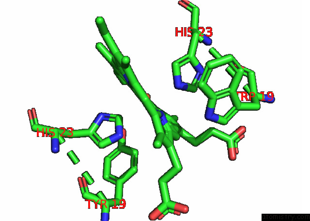
Mono view
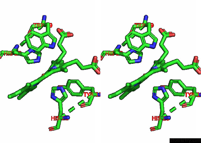
Stereo pair view
|
A full contact list of Iron with other atoms in the Fe binding
site number 2 of High-Resolution Structure of Photosystem II From the Mesophilic Cyanobacterium, Synechocystis Sp. Pcc 6803 within 5.0Å range:
probe
|
atom
|
residue
|
distance (Å)
|
B
|
Occ
|
F:Fe102
b:15.5
occ:1.00
|
FE
|
F:HEM102
|
0.0
|
15.5
|
1.0
|
NC
|
F:HEM102
|
2.0
|
15.5
|
1.0
|
NB
|
F:HEM102
|
2.1
|
15.5
|
1.0
|
ND
|
F:HEM102
|
2.1
|
15.5
|
1.0
|
NA
|
F:HEM102
|
2.1
|
15.5
|
1.0
|
NE2
|
F:HIS23
|
2.4
|
13.7
|
1.0
|
CE1
|
E:HIS23
|
2.5
|
15.5
|
1.0
|
C4D
|
F:HEM102
|
3.0
|
15.5
|
1.0
|
C1C
|
F:HEM102
|
3.0
|
15.5
|
1.0
|
C1A
|
F:HEM102
|
3.0
|
15.5
|
1.0
|
CD2
|
F:HIS23
|
3.0
|
13.7
|
1.0
|
C4B
|
F:HEM102
|
3.0
|
15.5
|
1.0
|
C1D
|
F:HEM102
|
3.1
|
15.5
|
1.0
|
C4A
|
F:HEM102
|
3.1
|
15.5
|
1.0
|
C1B
|
F:HEM102
|
3.1
|
15.5
|
1.0
|
C4C
|
F:HEM102
|
3.1
|
15.5
|
1.0
|
NE2
|
E:HIS23
|
3.2
|
15.5
|
1.0
|
CE1
|
F:HIS23
|
3.3
|
13.7
|
1.0
|
CHA
|
F:HEM102
|
3.4
|
15.5
|
1.0
|
CHC
|
F:HEM102
|
3.4
|
15.5
|
1.0
|
CHD
|
F:HEM102
|
3.4
|
15.5
|
1.0
|
CHB
|
F:HEM102
|
3.5
|
15.5
|
1.0
|
ND1
|
E:HIS23
|
3.5
|
15.5
|
1.0
|
CG
|
F:HIS23
|
4.1
|
13.7
|
1.0
|
C3D
|
F:HEM102
|
4.2
|
15.5
|
1.0
|
C2D
|
F:HEM102
|
4.2
|
15.5
|
1.0
|
C2A
|
F:HEM102
|
4.2
|
15.5
|
1.0
|
C3A
|
F:HEM102
|
4.2
|
15.5
|
1.0
|
ND1
|
F:HIS23
|
4.2
|
13.7
|
1.0
|
C2C
|
F:HEM102
|
4.3
|
15.5
|
1.0
|
C3B
|
F:HEM102
|
4.3
|
15.5
|
1.0
|
C2B
|
F:HEM102
|
4.3
|
15.5
|
1.0
|
C3C
|
F:HEM102
|
4.3
|
15.5
|
1.0
|
CD2
|
E:HIS23
|
4.5
|
15.5
|
1.0
|
CG
|
E:HIS23
|
4.6
|
15.5
|
1.0
|
NE1
|
F:TRP19
|
4.8
|
13.8
|
1.0
|
CE2
|
E:TYR19
|
4.8
|
15.9
|
1.0
|
|
Iron binding site 3 out
of 6 in 7rcv
Go back to
Iron Binding Sites List in 7rcv
Iron binding site 3 out
of 6 in the High-Resolution Structure of Photosystem II From the Mesophilic Cyanobacterium, Synechocystis Sp. Pcc 6803
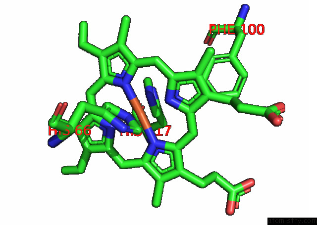
Mono view
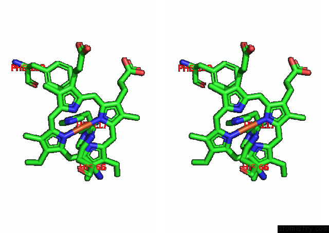
Stereo pair view
|
A full contact list of Iron with other atoms in the Fe binding
site number 3 of High-Resolution Structure of Photosystem II From the Mesophilic Cyanobacterium, Synechocystis Sp. Pcc 6803 within 5.0Å range:
probe
|
atom
|
residue
|
distance (Å)
|
B
|
Occ
|
V:Fe201
b:11.0
occ:1.00
|
FE
|
V:HEM201
|
0.0
|
11.0
|
1.0
|
ND
|
V:HEM201
|
2.0
|
11.0
|
1.0
|
NB
|
V:HEM201
|
2.0
|
11.0
|
1.0
|
NC
|
V:HEM201
|
2.0
|
11.0
|
1.0
|
NA
|
V:HEM201
|
2.0
|
11.0
|
1.0
|
NE2
|
V:HIS66
|
2.1
|
9.2
|
1.0
|
NE2
|
V:HIS117
|
2.4
|
11.1
|
1.0
|
CD2
|
V:HIS117
|
2.8
|
11.1
|
1.0
|
C4B
|
V:HEM201
|
3.0
|
11.0
|
1.0
|
C4D
|
V:HEM201
|
3.0
|
11.0
|
1.0
|
C1D
|
V:HEM201
|
3.0
|
11.0
|
1.0
|
C1B
|
V:HEM201
|
3.0
|
11.0
|
1.0
|
C1C
|
V:HEM201
|
3.0
|
11.0
|
1.0
|
C1A
|
V:HEM201
|
3.0
|
11.0
|
1.0
|
C4C
|
V:HEM201
|
3.1
|
11.0
|
1.0
|
C4A
|
V:HEM201
|
3.1
|
11.0
|
1.0
|
CD2
|
V:HIS66
|
3.1
|
9.2
|
1.0
|
CE1
|
V:HIS66
|
3.2
|
9.2
|
1.0
|
CHC
|
V:HEM201
|
3.4
|
11.0
|
1.0
|
CHA
|
V:HEM201
|
3.4
|
11.0
|
1.0
|
CHD
|
V:HEM201
|
3.4
|
11.0
|
1.0
|
CHB
|
V:HEM201
|
3.4
|
11.0
|
1.0
|
CE1
|
V:HIS117
|
3.6
|
11.1
|
1.0
|
CG
|
V:HIS117
|
4.1
|
11.1
|
1.0
|
C3B
|
V:HEM201
|
4.2
|
11.0
|
1.0
|
C2B
|
V:HEM201
|
4.2
|
11.0
|
1.0
|
C3D
|
V:HEM201
|
4.2
|
11.0
|
1.0
|
C2D
|
V:HEM201
|
4.2
|
11.0
|
1.0
|
ND1
|
V:HIS66
|
4.3
|
9.2
|
1.0
|
CG
|
V:HIS66
|
4.3
|
9.2
|
1.0
|
C2C
|
V:HEM201
|
4.3
|
11.0
|
1.0
|
C3C
|
V:HEM201
|
4.3
|
11.0
|
1.0
|
C2A
|
V:HEM201
|
4.3
|
11.0
|
1.0
|
C3A
|
V:HEM201
|
4.3
|
11.0
|
1.0
|
ND1
|
V:HIS117
|
4.5
|
11.1
|
1.0
|
CE1
|
V:PHE100
|
4.9
|
13.4
|
1.0
|
|
Iron binding site 4 out
of 6 in 7rcv
Go back to
Iron Binding Sites List in 7rcv
Iron binding site 4 out
of 6 in the High-Resolution Structure of Photosystem II From the Mesophilic Cyanobacterium, Synechocystis Sp. Pcc 6803
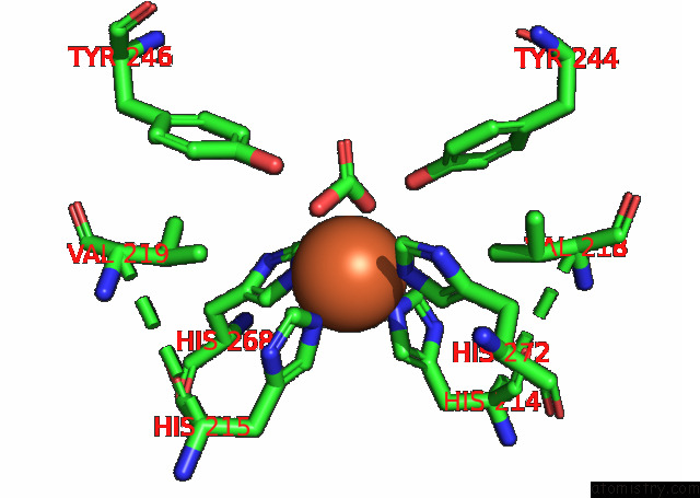
Mono view
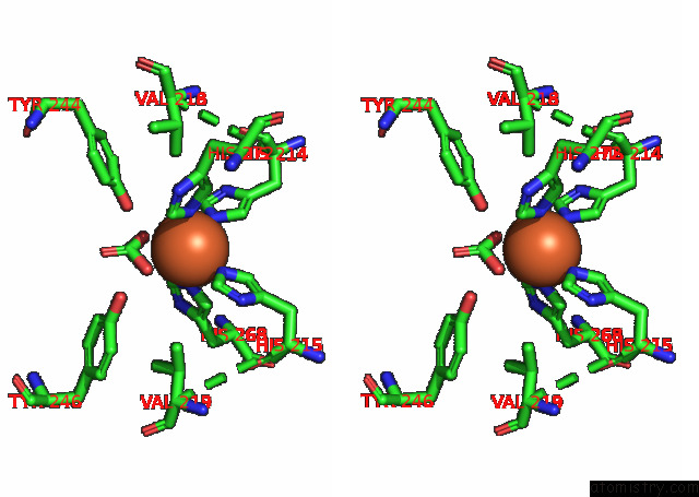
Stereo pair view
|
A full contact list of Iron with other atoms in the Fe binding
site number 4 of High-Resolution Structure of Photosystem II From the Mesophilic Cyanobacterium, Synechocystis Sp. Pcc 6803 within 5.0Å range:
probe
|
atom
|
residue
|
distance (Å)
|
B
|
Occ
|
a:Fe402
b:3.3
occ:1.00
|
NE2
|
a:HIS215
|
2.3
|
3.6
|
1.0
|
O1
|
a:BCT418
|
2.4
|
4.2
|
1.0
|
O3
|
a:BCT418
|
2.4
|
4.2
|
1.0
|
NE2
|
d:HIS268
|
2.4
|
3.7
|
1.0
|
NE2
|
a:HIS272
|
2.4
|
3.5
|
1.0
|
NE2
|
d:HIS214
|
2.4
|
10.5
|
1.0
|
C
|
a:BCT418
|
2.8
|
4.2
|
1.0
|
CD2
|
a:HIS215
|
3.2
|
3.6
|
1.0
|
CD2
|
d:HIS214
|
3.3
|
10.5
|
1.0
|
CE1
|
a:HIS272
|
3.3
|
3.5
|
1.0
|
CD2
|
d:HIS268
|
3.3
|
3.7
|
1.0
|
CE1
|
a:HIS215
|
3.3
|
3.6
|
1.0
|
CE1
|
d:HIS268
|
3.4
|
3.7
|
1.0
|
CD2
|
a:HIS272
|
3.4
|
3.5
|
1.0
|
CE1
|
d:HIS214
|
3.5
|
10.5
|
1.0
|
O2
|
a:BCT418
|
4.1
|
4.2
|
1.0
|
CG
|
a:HIS215
|
4.4
|
3.6
|
1.0
|
ND1
|
a:HIS215
|
4.4
|
3.6
|
1.0
|
ND1
|
a:HIS272
|
4.5
|
3.5
|
1.0
|
CG
|
d:HIS214
|
4.5
|
10.5
|
1.0
|
OH
|
d:TYR244
|
4.5
|
4.3
|
1.0
|
CG
|
d:HIS268
|
4.5
|
3.7
|
1.0
|
ND1
|
d:HIS268
|
4.5
|
3.7
|
1.0
|
ND1
|
d:HIS214
|
4.5
|
10.5
|
1.0
|
CG
|
a:HIS272
|
4.6
|
3.5
|
1.0
|
CG2
|
a:VAL219
|
4.7
|
3.1
|
1.0
|
OH
|
a:TYR246
|
4.7
|
8.6
|
1.0
|
CG2
|
d:VAL218
|
4.8
|
2.6
|
1.0
|
|
Iron binding site 5 out
of 6 in 7rcv
Go back to
Iron Binding Sites List in 7rcv
Iron binding site 5 out
of 6 in the High-Resolution Structure of Photosystem II From the Mesophilic Cyanobacterium, Synechocystis Sp. Pcc 6803
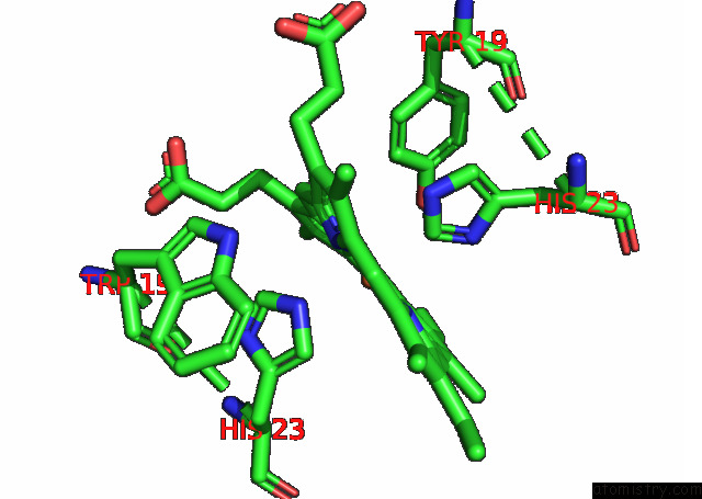
Mono view
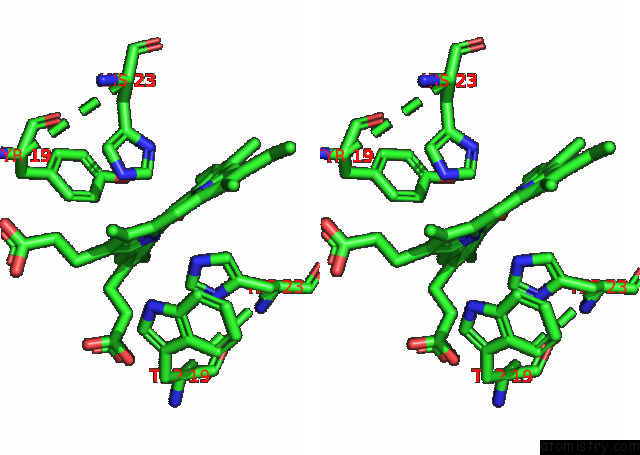
Stereo pair view
|
A full contact list of Iron with other atoms in the Fe binding
site number 5 of High-Resolution Structure of Photosystem II From the Mesophilic Cyanobacterium, Synechocystis Sp. Pcc 6803 within 5.0Å range:
probe
|
atom
|
residue
|
distance (Å)
|
B
|
Occ
|
f:Fe102
b:15.5
occ:1.00
|
FE
|
f:HEM102
|
0.0
|
15.5
|
1.0
|
NC
|
f:HEM102
|
2.0
|
15.5
|
1.0
|
NB
|
f:HEM102
|
2.1
|
15.5
|
1.0
|
ND
|
f:HEM102
|
2.1
|
15.5
|
1.0
|
NA
|
f:HEM102
|
2.1
|
15.5
|
1.0
|
NE2
|
f:HIS23
|
2.4
|
13.7
|
1.0
|
CE1
|
e:HIS23
|
2.5
|
15.5
|
1.0
|
C4D
|
f:HEM102
|
3.0
|
15.5
|
1.0
|
C1C
|
f:HEM102
|
3.0
|
15.5
|
1.0
|
C1A
|
f:HEM102
|
3.0
|
15.5
|
1.0
|
CD2
|
f:HIS23
|
3.0
|
13.7
|
1.0
|
C4B
|
f:HEM102
|
3.0
|
15.5
|
1.0
|
C1D
|
f:HEM102
|
3.1
|
15.5
|
1.0
|
C4A
|
f:HEM102
|
3.1
|
15.5
|
1.0
|
C1B
|
f:HEM102
|
3.1
|
15.5
|
1.0
|
C4C
|
f:HEM102
|
3.1
|
15.5
|
1.0
|
NE2
|
e:HIS23
|
3.2
|
15.5
|
1.0
|
CE1
|
f:HIS23
|
3.3
|
13.7
|
1.0
|
CHA
|
f:HEM102
|
3.4
|
15.5
|
1.0
|
CHC
|
f:HEM102
|
3.4
|
15.5
|
1.0
|
CHD
|
f:HEM102
|
3.4
|
15.5
|
1.0
|
CHB
|
f:HEM102
|
3.5
|
15.5
|
1.0
|
ND1
|
e:HIS23
|
3.5
|
15.5
|
1.0
|
CG
|
f:HIS23
|
4.1
|
13.7
|
1.0
|
C3D
|
f:HEM102
|
4.2
|
15.5
|
1.0
|
C2D
|
f:HEM102
|
4.2
|
15.5
|
1.0
|
C2A
|
f:HEM102
|
4.2
|
15.5
|
1.0
|
C3A
|
f:HEM102
|
4.2
|
15.5
|
1.0
|
ND1
|
f:HIS23
|
4.2
|
13.7
|
1.0
|
C2C
|
f:HEM102
|
4.2
|
15.5
|
1.0
|
C3B
|
f:HEM102
|
4.3
|
15.5
|
1.0
|
C2B
|
f:HEM102
|
4.3
|
15.5
|
1.0
|
C3C
|
f:HEM102
|
4.3
|
15.5
|
1.0
|
CD2
|
e:HIS23
|
4.5
|
15.5
|
1.0
|
CG
|
e:HIS23
|
4.6
|
15.5
|
1.0
|
NE1
|
f:TRP19
|
4.8
|
13.8
|
1.0
|
CE2
|
e:TYR19
|
4.8
|
15.9
|
1.0
|
|
Iron binding site 6 out
of 6 in 7rcv
Go back to
Iron Binding Sites List in 7rcv
Iron binding site 6 out
of 6 in the High-Resolution Structure of Photosystem II From the Mesophilic Cyanobacterium, Synechocystis Sp. Pcc 6803
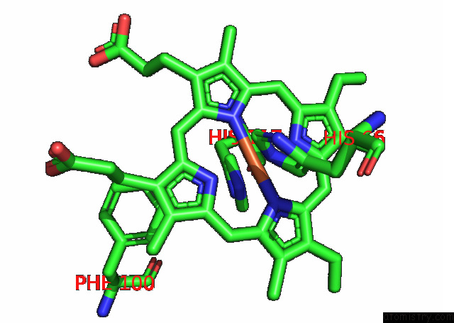
Mono view
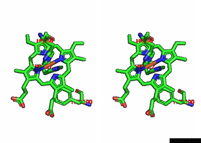
Stereo pair view
|
A full contact list of Iron with other atoms in the Fe binding
site number 6 of High-Resolution Structure of Photosystem II From the Mesophilic Cyanobacterium, Synechocystis Sp. Pcc 6803 within 5.0Å range:
probe
|
atom
|
residue
|
distance (Å)
|
B
|
Occ
|
v:Fe201
b:11.0
occ:1.00
|
FE
|
v:HEM201
|
0.0
|
11.0
|
1.0
|
ND
|
v:HEM201
|
2.0
|
11.0
|
1.0
|
NB
|
v:HEM201
|
2.0
|
11.0
|
1.0
|
NA
|
v:HEM201
|
2.0
|
11.0
|
1.0
|
NC
|
v:HEM201
|
2.0
|
11.0
|
1.0
|
NE2
|
v:HIS66
|
2.1
|
9.2
|
1.0
|
NE2
|
v:HIS117
|
2.4
|
11.1
|
1.0
|
CD2
|
v:HIS117
|
2.8
|
11.1
|
1.0
|
C4B
|
v:HEM201
|
3.0
|
11.0
|
1.0
|
C4D
|
v:HEM201
|
3.0
|
11.0
|
1.0
|
C1D
|
v:HEM201
|
3.0
|
11.0
|
1.0
|
C1B
|
v:HEM201
|
3.0
|
11.0
|
1.0
|
C1C
|
v:HEM201
|
3.0
|
11.0
|
1.0
|
C1A
|
v:HEM201
|
3.0
|
11.0
|
1.0
|
C4C
|
v:HEM201
|
3.1
|
11.0
|
1.0
|
C4A
|
v:HEM201
|
3.1
|
11.0
|
1.0
|
CD2
|
v:HIS66
|
3.1
|
9.2
|
1.0
|
CE1
|
v:HIS66
|
3.2
|
9.2
|
1.0
|
CHC
|
v:HEM201
|
3.4
|
11.0
|
1.0
|
CHA
|
v:HEM201
|
3.4
|
11.0
|
1.0
|
CHD
|
v:HEM201
|
3.4
|
11.0
|
1.0
|
CHB
|
v:HEM201
|
3.4
|
11.0
|
1.0
|
CE1
|
v:HIS117
|
3.6
|
11.1
|
1.0
|
CG
|
v:HIS117
|
4.1
|
11.1
|
1.0
|
C3B
|
v:HEM201
|
4.2
|
11.0
|
1.0
|
C2B
|
v:HEM201
|
4.2
|
11.0
|
1.0
|
C3D
|
v:HEM201
|
4.2
|
11.0
|
1.0
|
C2D
|
v:HEM201
|
4.2
|
11.0
|
1.0
|
ND1
|
v:HIS66
|
4.3
|
9.2
|
1.0
|
C2C
|
v:HEM201
|
4.3
|
11.0
|
1.0
|
CG
|
v:HIS66
|
4.3
|
9.2
|
1.0
|
C3C
|
v:HEM201
|
4.3
|
11.0
|
1.0
|
C2A
|
v:HEM201
|
4.3
|
11.0
|
1.0
|
C3A
|
v:HEM201
|
4.3
|
11.0
|
1.0
|
ND1
|
v:HIS117
|
4.5
|
11.1
|
1.0
|
CE1
|
v:PHE100
|
4.9
|
13.4
|
1.0
|
|
Reference:
C.J.Gisriel,
J.Wang,
J.Liu,
D.A.Flesher,
K.M.Reiss,
H.Huang,
K.R.Yang,
W.H.Armstrong,
M.R.Gunner,
V.S.Batista,
R.J.Debus,
G.W.Brudvig.
High-Resolution Cryo-Em Structure of Photosystem II From the Mesophilic Cyanobacterium, Synechocystis Sp. Pcc 6803 To Be Published.
Page generated: Thu Aug 8 23:40:15 2024
|