Iron »
PDB 7sh5-7tc8 »
7snm »
Iron in PDB 7snm: Lanosterol-Bound P450 Domain of the CYP51-Ferredoxin Fusion Protein From Methylococcus Capsulatus
Enzymatic activity of Lanosterol-Bound P450 Domain of the CYP51-Ferredoxin Fusion Protein From Methylococcus Capsulatus
All present enzymatic activity of Lanosterol-Bound P450 Domain of the CYP51-Ferredoxin Fusion Protein From Methylococcus Capsulatus:
1.14.13.70;
1.14.13.70;
Protein crystallography data
The structure of Lanosterol-Bound P450 Domain of the CYP51-Ferredoxin Fusion Protein From Methylococcus Capsulatus, PDB code: 7snm
was solved by
G.I.Lepesheva,
T.Hargrove,
Z.Wawrzak,
with X-Ray Crystallography technique. A brief refinement statistics is given in the table below:
| Resolution Low / High (Å) | 74.45 / 2.55 |
| Space group | P 1 21 1 |
| Cell size a, b, c (Å), α, β, γ (°) | 73.21, 206.16, 81.43, 90, 114.09, 90 |
| R / Rfree (%) | 20.5 / 24.6 |
Iron Binding Sites:
The binding sites of Iron atom in the Lanosterol-Bound P450 Domain of the CYP51-Ferredoxin Fusion Protein From Methylococcus Capsulatus
(pdb code 7snm). This binding sites where shown within
5.0 Angstroms radius around Iron atom.
In total 4 binding sites of Iron where determined in the Lanosterol-Bound P450 Domain of the CYP51-Ferredoxin Fusion Protein From Methylococcus Capsulatus, PDB code: 7snm:
Jump to Iron binding site number: 1; 2; 3; 4;
In total 4 binding sites of Iron where determined in the Lanosterol-Bound P450 Domain of the CYP51-Ferredoxin Fusion Protein From Methylococcus Capsulatus, PDB code: 7snm:
Jump to Iron binding site number: 1; 2; 3; 4;
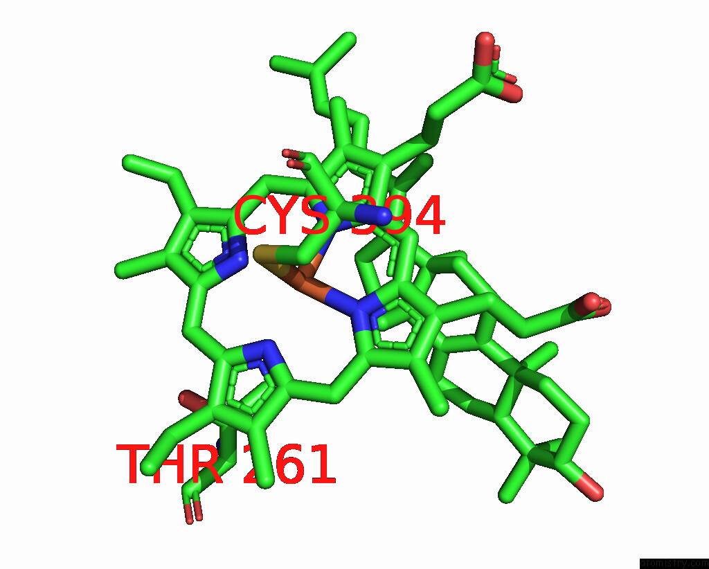
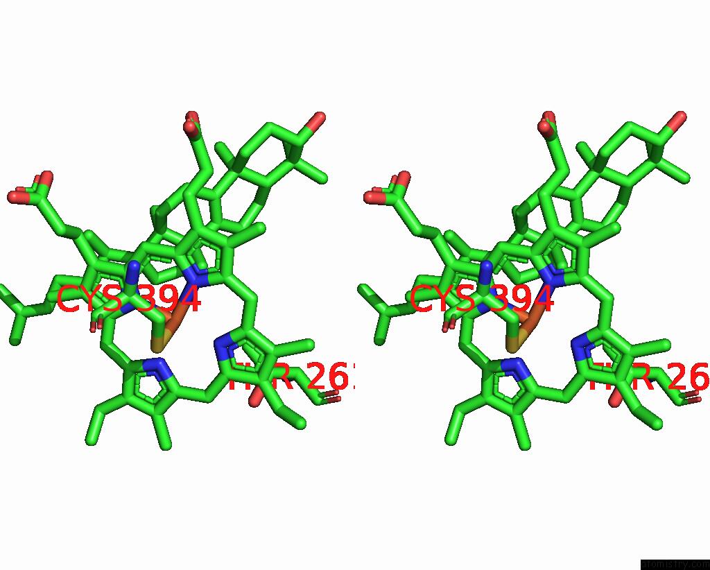
Iron binding site 1 out of 4 in 7snm
Go back to
Iron binding site 1 out
of 4 in the Lanosterol-Bound P450 Domain of the CYP51-Ferredoxin Fusion Protein From Methylococcus Capsulatus
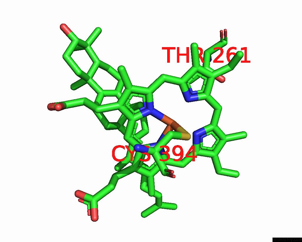
Mono view
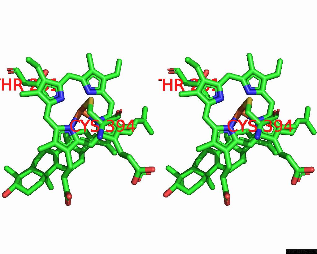
Stereo pair view
Mono view
Stereo pair view
A full contact list of Iron with other atoms in the Fe binding
site number 1 of Lanosterol-Bound P450 Domain of the CYP51-Ferredoxin Fusion Protein From Methylococcus Capsulatus within 5.0Å range:
|
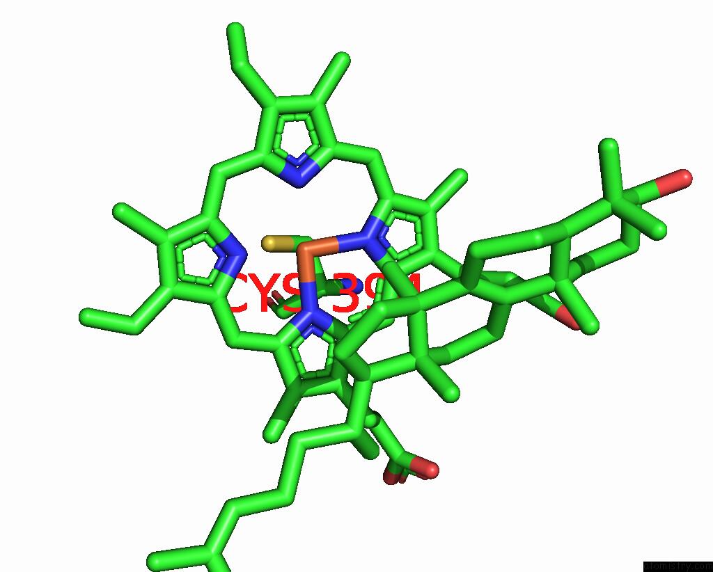
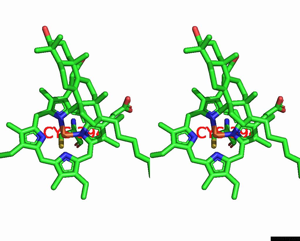
Iron binding site 2 out of 4 in 7snm
Go back to
Iron binding site 2 out
of 4 in the Lanosterol-Bound P450 Domain of the CYP51-Ferredoxin Fusion Protein From Methylococcus Capsulatus
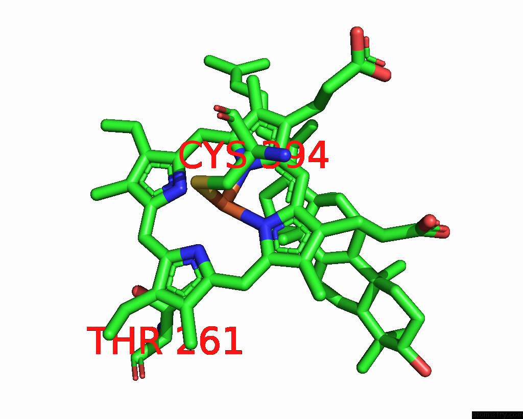
Mono view
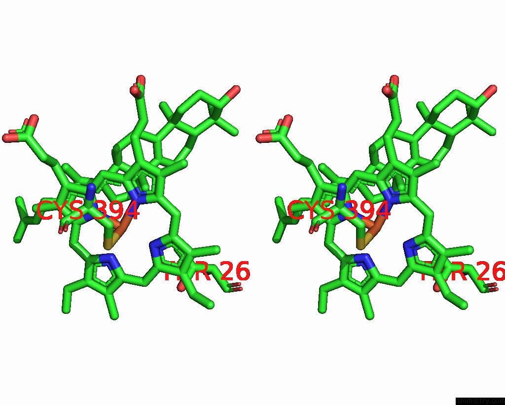
Stereo pair view
Mono view
Stereo pair view
A full contact list of Iron with other atoms in the Fe binding
site number 2 of Lanosterol-Bound P450 Domain of the CYP51-Ferredoxin Fusion Protein From Methylococcus Capsulatus within 5.0Å range:
|
Iron binding site 3 out of 4 in 7snm
Go back to
Iron binding site 3 out
of 4 in the Lanosterol-Bound P450 Domain of the CYP51-Ferredoxin Fusion Protein From Methylococcus Capsulatus
Mono view
Stereo pair view
Mono view
Stereo pair view
A full contact list of Iron with other atoms in the Fe binding
site number 3 of Lanosterol-Bound P450 Domain of the CYP51-Ferredoxin Fusion Protein From Methylococcus Capsulatus within 5.0Å range:
|
Iron binding site 4 out of 4 in 7snm
Go back to
Iron binding site 4 out
of 4 in the Lanosterol-Bound P450 Domain of the CYP51-Ferredoxin Fusion Protein From Methylococcus Capsulatus
Mono view
Stereo pair view
Mono view
Stereo pair view
A full contact list of Iron with other atoms in the Fe binding
site number 4 of Lanosterol-Bound P450 Domain of the CYP51-Ferredoxin Fusion Protein From Methylococcus Capsulatus within 5.0Å range:
|
Reference:
T.Y.Hargrove,
D.C.Lamb,
J.A.Smith,
Z.Wawrzak,
S.L.Kelly,
G.I.Lepesheva.
Unravelling the Role of Transient Redox Partner Complexes in P450 Electron Transfer Mechanics. Sci Rep V. 12 16232 2022.
ISSN: ESSN 2045-2322
PubMed: 36171457
DOI: 10.1038/S41598-022-20671-0
Page generated: Thu Aug 7 05:34:57 2025
ISSN: ESSN 2045-2322
PubMed: 36171457
DOI: 10.1038/S41598-022-20671-0
Last articles
Fe in 7VYPFe in 7VYO
Fe in 7VYJ
Fe in 7VYA
Fe in 7VY8
Fe in 7VT2
Fe in 7VXU
Fe in 7VY3
Fe in 7VXP
Fe in 7VXQ