Iron »
PDB 7tce-7tqh »
7tl2 »
Iron in PDB 7tl2: Crystal Structure of Yeast P58C Multi-Tyrosine Mutant 5YF412
Protein crystallography data
The structure of Crystal Structure of Yeast P58C Multi-Tyrosine Mutant 5YF412, PDB code: 7tl2
was solved by
A.M.Blee,
L.E.Salay,
W.J.Chazin,
with X-Ray Crystallography technique. A brief refinement statistics is given in the table below:
| Resolution Low / High (Å) | 26.02 / 1.53 |
| Space group | P 21 21 21 |
| Cell size a, b, c (Å), α, β, γ (°) | 40.624, 51.87, 89.479, 90, 90, 90 |
| R / Rfree (%) | 15.2 / 16.8 |
Iron Binding Sites:
The binding sites of Iron atom in the Crystal Structure of Yeast P58C Multi-Tyrosine Mutant 5YF412
(pdb code 7tl2). This binding sites where shown within
5.0 Angstroms radius around Iron atom.
In total 4 binding sites of Iron where determined in the Crystal Structure of Yeast P58C Multi-Tyrosine Mutant 5YF412, PDB code: 7tl2:
Jump to Iron binding site number: 1; 2; 3; 4;
In total 4 binding sites of Iron where determined in the Crystal Structure of Yeast P58C Multi-Tyrosine Mutant 5YF412, PDB code: 7tl2:
Jump to Iron binding site number: 1; 2; 3; 4;
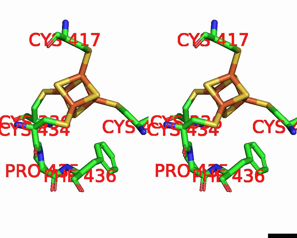
Iron binding site 1 out of 4 in 7tl2
Go back to
Iron binding site 1 out
of 4 in the Crystal Structure of Yeast P58C Multi-Tyrosine Mutant 5YF412

Mono view
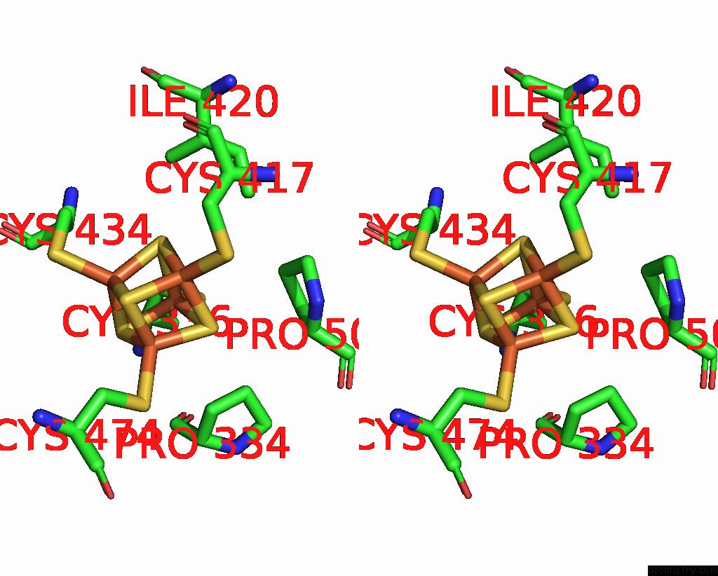
Stereo pair view
Mono view
Stereo pair view
A full contact list of Iron with other atoms in the Fe binding
site number 1 of Crystal Structure of Yeast P58C Multi-Tyrosine Mutant 5YF412 within 5.0Å range:
|
Iron binding site 2 out of 4 in 7tl2
Go back to
Iron binding site 2 out
of 4 in the Crystal Structure of Yeast P58C Multi-Tyrosine Mutant 5YF412
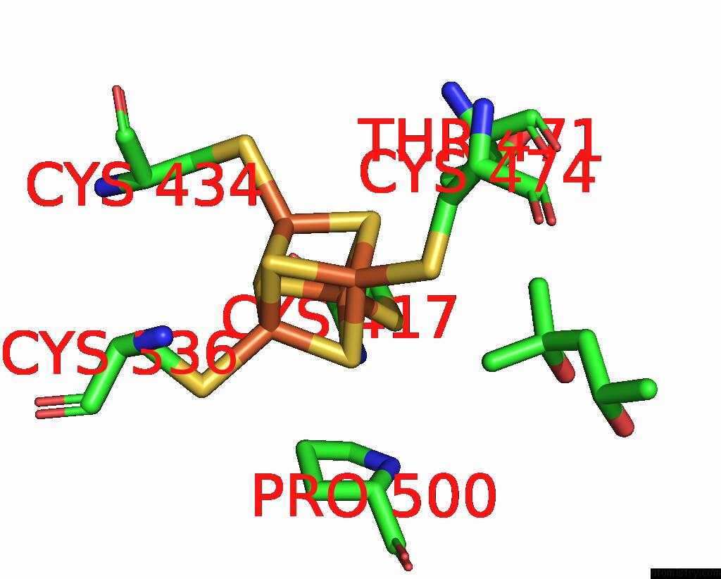
Mono view

Stereo pair view
Mono view
Stereo pair view
A full contact list of Iron with other atoms in the Fe binding
site number 2 of Crystal Structure of Yeast P58C Multi-Tyrosine Mutant 5YF412 within 5.0Å range:
|
Iron binding site 3 out of 4 in 7tl2
Go back to
Iron binding site 3 out
of 4 in the Crystal Structure of Yeast P58C Multi-Tyrosine Mutant 5YF412
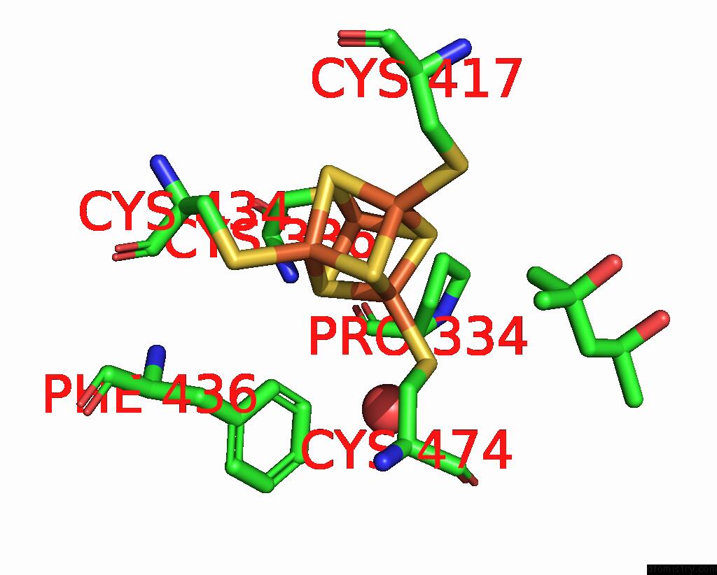
Mono view
Stereo pair view
Mono view
Stereo pair view
A full contact list of Iron with other atoms in the Fe binding
site number 3 of Crystal Structure of Yeast P58C Multi-Tyrosine Mutant 5YF412 within 5.0Å range:
|
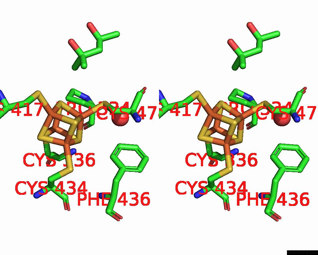
Iron binding site 4 out of 4 in 7tl2
Go back to
Iron binding site 4 out
of 4 in the Crystal Structure of Yeast P58C Multi-Tyrosine Mutant 5YF412

Mono view
Stereo pair view
Mono view
Stereo pair view
A full contact list of Iron with other atoms in the Fe binding
site number 4 of Crystal Structure of Yeast P58C Multi-Tyrosine Mutant 5YF412 within 5.0Å range:
|
Reference:
L.E.Salay,
A.M.Blee,
M.K.Raza,
K.S.Gallagher,
H.Chen,
A.J.Dorfeuille,
J.K.Barton,
W.J.Chazin.
Modification of the 4FE-4S Cluster Charge Transport Pathway Alters Rna Synthesis By Yeast Dna Primase. Biochemistry V. 61 1113 2022.
ISSN: ISSN 0006-2960
PubMed: 35617695
DOI: 10.1021/ACS.BIOCHEM.2C00100
Page generated: Thu Aug 7 05:56:57 2025
ISSN: ISSN 0006-2960
PubMed: 35617695
DOI: 10.1021/ACS.BIOCHEM.2C00100
Last articles
Mg in 3J7HMg in 3J7I
Mg in 3IVK
Mg in 3J6E
Mg in 3J6G
Mg in 3J6P
Mg in 3J6H
Mg in 3J1F
Mg in 3J6F
Mg in 3J5V