Iron »
PDB 7tc8-7tqf »
7tl3 »
Iron in PDB 7tl3: Crystal Structure of Yeast P58C Multi-Tyrosine Mutant 5YF431
Protein crystallography data
The structure of Crystal Structure of Yeast P58C Multi-Tyrosine Mutant 5YF431, PDB code: 7tl3
was solved by
A.M.Blee,
L.E.Salay,
W.J.Chazin,
with X-Ray Crystallography technique. A brief refinement statistics is given in the table below:
| Resolution Low / High (Å) | 30.36 / 2.07 |
| Space group | P 21 21 21 |
| Cell size a, b, c (Å), α, β, γ (°) | 41.044, 51.097, 90.223, 90, 90, 90 |
| R / Rfree (%) | 23.6 / 27.7 |
Iron Binding Sites:
The binding sites of Iron atom in the Crystal Structure of Yeast P58C Multi-Tyrosine Mutant 5YF431
(pdb code 7tl3). This binding sites where shown within
5.0 Angstroms radius around Iron atom.
In total 4 binding sites of Iron where determined in the Crystal Structure of Yeast P58C Multi-Tyrosine Mutant 5YF431, PDB code: 7tl3:
Jump to Iron binding site number: 1; 2; 3; 4;
In total 4 binding sites of Iron where determined in the Crystal Structure of Yeast P58C Multi-Tyrosine Mutant 5YF431, PDB code: 7tl3:
Jump to Iron binding site number: 1; 2; 3; 4;
Iron binding site 1 out of 4 in 7tl3
Go back to
Iron binding site 1 out
of 4 in the Crystal Structure of Yeast P58C Multi-Tyrosine Mutant 5YF431

Mono view
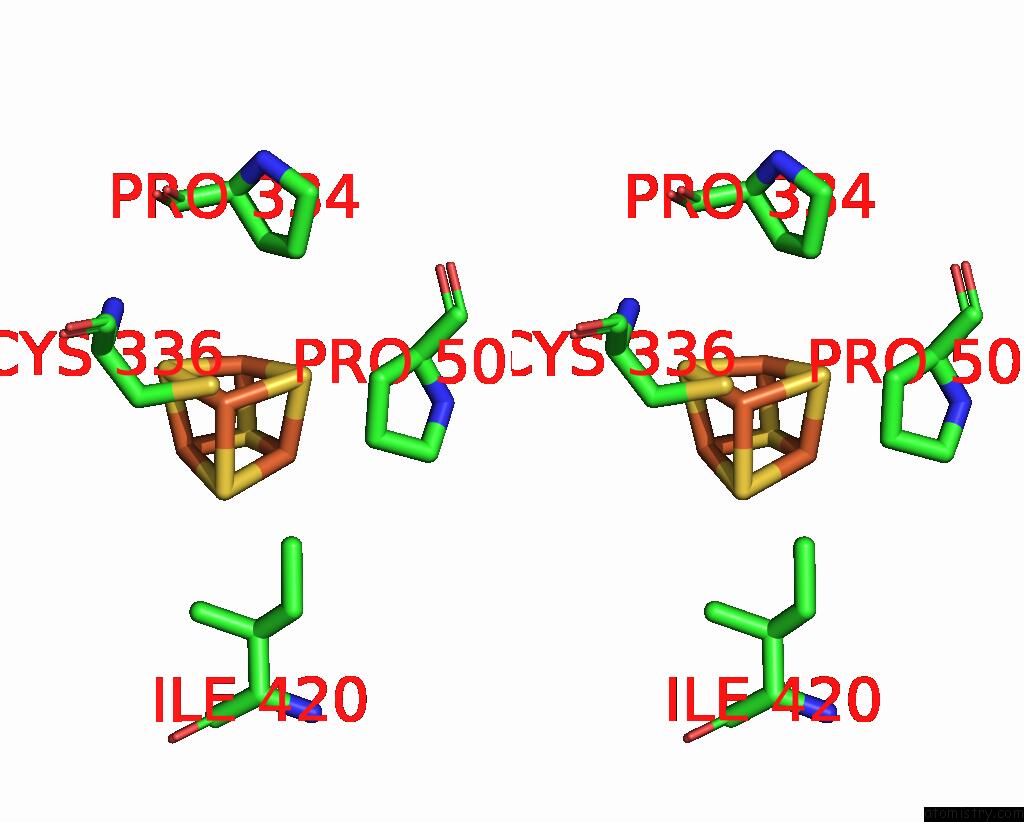
Stereo pair view
Mono view
Stereo pair view
A full contact list of Iron with other atoms in the Fe binding
site number 1 of Crystal Structure of Yeast P58C Multi-Tyrosine Mutant 5YF431 within 5.0Å range:
|
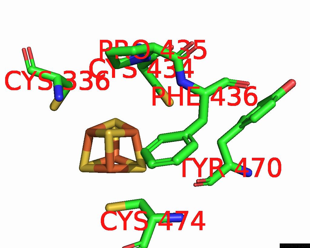
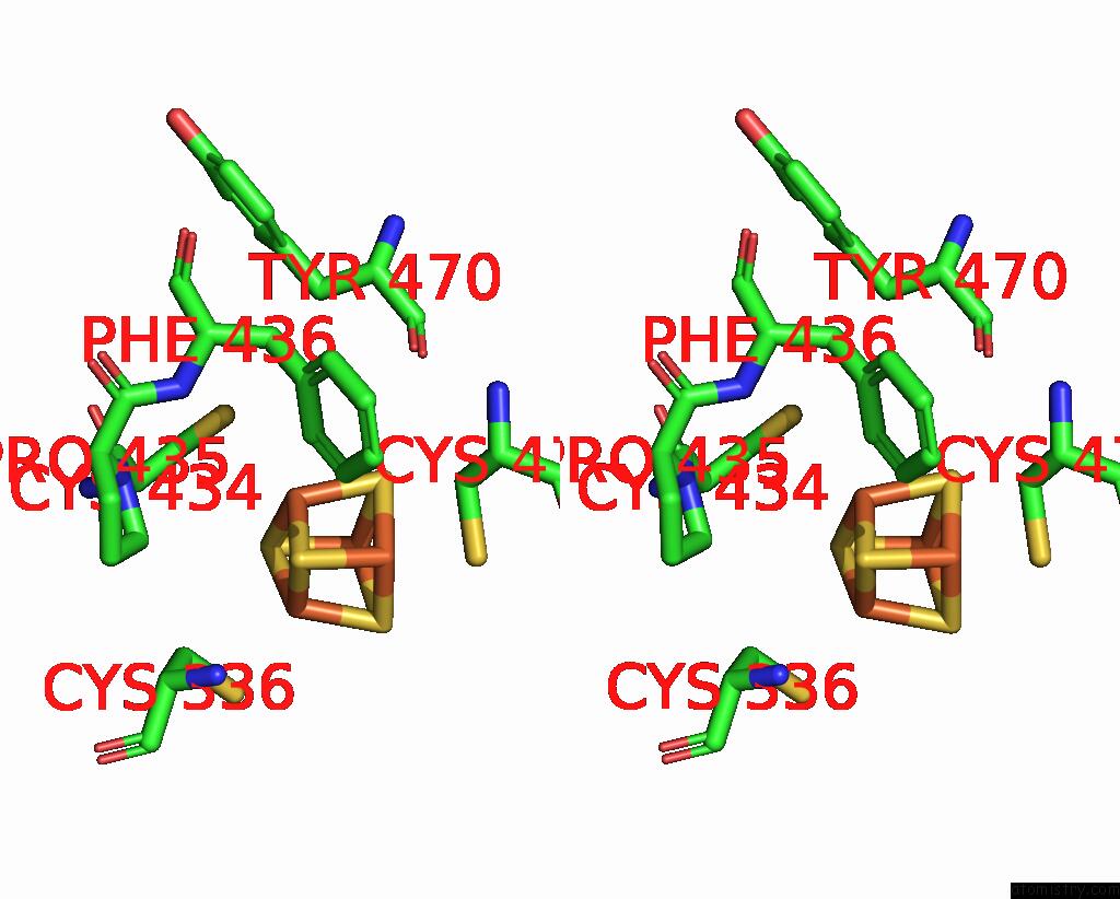
Iron binding site 2 out of 4 in 7tl3
Go back to
Iron binding site 2 out
of 4 in the Crystal Structure of Yeast P58C Multi-Tyrosine Mutant 5YF431

Mono view

Stereo pair view
Mono view
Stereo pair view
A full contact list of Iron with other atoms in the Fe binding
site number 2 of Crystal Structure of Yeast P58C Multi-Tyrosine Mutant 5YF431 within 5.0Å range:
|
Iron binding site 3 out of 4 in 7tl3
Go back to
Iron binding site 3 out
of 4 in the Crystal Structure of Yeast P58C Multi-Tyrosine Mutant 5YF431

Mono view

Stereo pair view
Mono view
Stereo pair view
A full contact list of Iron with other atoms in the Fe binding
site number 3 of Crystal Structure of Yeast P58C Multi-Tyrosine Mutant 5YF431 within 5.0Å range:
|
Iron binding site 4 out of 4 in 7tl3
Go back to
Iron binding site 4 out
of 4 in the Crystal Structure of Yeast P58C Multi-Tyrosine Mutant 5YF431
Mono view
Stereo pair view
Mono view
Stereo pair view
A full contact list of Iron with other atoms in the Fe binding
site number 4 of Crystal Structure of Yeast P58C Multi-Tyrosine Mutant 5YF431 within 5.0Å range:
|
Reference:
L.E.Salay,
A.M.Blee,
M.K.Raza,
K.S.Gallagher,
H.Chen,
A.J.Dorfeuille,
J.K.Barton,
W.J.Chazin.
Modification of the 4FE-4S Cluster Charge Transport Pathway Alters Rna Synthesis By Yeast Dna Primase. Biochemistry V. 61 1113 2022.
ISSN: ISSN 0006-2960
PubMed: 35617695
DOI: 10.1021/ACS.BIOCHEM.2C00100
Page generated: Fri Aug 9 01:56:37 2024
ISSN: ISSN 0006-2960
PubMed: 35617695
DOI: 10.1021/ACS.BIOCHEM.2C00100
Last articles
F in 7PVKF in 7Q2J
F in 7Q01
F in 7PZX
F in 7PZW
F in 7PZV
F in 7PZU
F in 7PZS
F in 7PY4
F in 7PX6