Iron »
PDB 7tce-7tqh »
7tlx »
Iron in PDB 7tlx: Crystal Structure of Cytochrome C From Pseudomonas Putida S16
Protein crystallography data
The structure of Crystal Structure of Cytochrome C From Pseudomonas Putida S16, PDB code: 7tlx
was solved by
K.Wu,
M.Dulchavsky,
F.Stull,
J.C.A.Bardwell,
with X-Ray Crystallography technique. A brief refinement statistics is given in the table below:
| Resolution Low / High (Å) | 41.84 / 1.90 |
| Space group | C 1 2 1 |
| Cell size a, b, c (Å), α, β, γ (°) | 79.73, 30.17, 49.1, 90, 121.56, 90 |
| R / Rfree (%) | 15.1 / 17.5 |
Iron Binding Sites:
The binding sites of Iron atom in the Crystal Structure of Cytochrome C From Pseudomonas Putida S16
(pdb code 7tlx). This binding sites where shown within
5.0 Angstroms radius around Iron atom.
In total only one binding site of Iron was determined in the Crystal Structure of Cytochrome C From Pseudomonas Putida S16, PDB code: 7tlx:
In total only one binding site of Iron was determined in the Crystal Structure of Cytochrome C From Pseudomonas Putida S16, PDB code: 7tlx:
Iron binding site 1 out of 1 in 7tlx
Go back to
Iron binding site 1 out
of 1 in the Crystal Structure of Cytochrome C From Pseudomonas Putida S16

Mono view
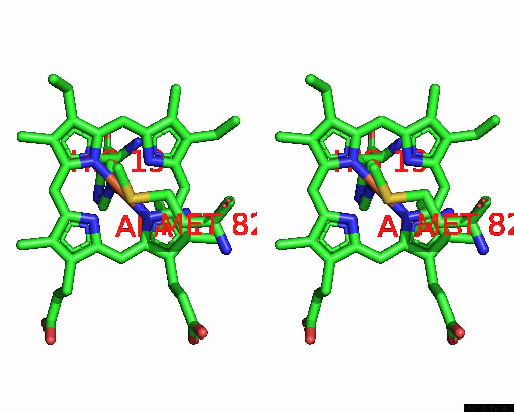
Stereo pair view
Mono view
Stereo pair view
A full contact list of Iron with other atoms in the Fe binding
site number 1 of Crystal Structure of Cytochrome C From Pseudomonas Putida S16 within 5.0Å range:
|
Reference:
V.Choudhary,
K.Wu,
Z.Zhang,
M.Dulchavsky,
T.Barkman,
J.C.A.Bardwell,
F.Stull.
The Enzyme Pseudooxynicotine Amine Oxidase From Pseudomonas Putida S16 Is Not An Oxidase, But A Dehydrogenase. J.Biol.Chem. V. 298 02251 2022.
ISSN: ESSN 1083-351X
PubMed: 35835223
DOI: 10.1016/J.JBC.2022.102251
Page generated: Thu Aug 7 05:58:55 2025
ISSN: ESSN 1083-351X
PubMed: 35835223
DOI: 10.1016/J.JBC.2022.102251
Last articles
Mg in 2A6EMg in 2A5Z
Mg in 2A5L
Mg in 2A5Y
Mg in 2A5J
Mg in 2A43
Mg in 2A5G
Mg in 2A5D
Mg in 2A5F
Mg in 2A42