Iron »
PDB 7tc8-7tqf »
7tom »
Iron in PDB 7tom: X-Ray Crystal Structure of Glycerol Dibiphytanyl Glycerol Tetraether - Macrocyclic Archaeol Synthase (Gdgt-Mas) From Methanocaldococcus Jannaschii with Bacterial Lipid Substrate Analog, 5'Deoxyadenosine, and Methionine Bound
Protein crystallography data
The structure of X-Ray Crystal Structure of Glycerol Dibiphytanyl Glycerol Tetraether - Macrocyclic Archaeol Synthase (Gdgt-Mas) From Methanocaldococcus Jannaschii with Bacterial Lipid Substrate Analog, 5'Deoxyadenosine, and Methionine Bound, PDB code: 7tom
was solved by
C.T.Lloyd,
S.J.Booker,
A.K.Boal,
with X-Ray Crystallography technique. A brief refinement statistics is given in the table below:
| Resolution Low / High (Å) | 45.54 / 1.85 |
| Space group | P 21 21 21 |
| Cell size a, b, c (Å), α, β, γ (°) | 55.609, 77.158, 112.817, 90, 90, 90 |
| R / Rfree (%) | 15 / 19.9 |
Iron Binding Sites:
Pages:
>>> Page 1 <<< Page 2, Binding sites: 11 - 13;Binding sites:
The binding sites of Iron atom in the X-Ray Crystal Structure of Glycerol Dibiphytanyl Glycerol Tetraether - Macrocyclic Archaeol Synthase (Gdgt-Mas) From Methanocaldococcus Jannaschii with Bacterial Lipid Substrate Analog, 5'Deoxyadenosine, and Methionine Bound (pdb code 7tom). This binding sites where shown within 5.0 Angstroms radius around Iron atom.In total 13 binding sites of Iron where determined in the X-Ray Crystal Structure of Glycerol Dibiphytanyl Glycerol Tetraether - Macrocyclic Archaeol Synthase (Gdgt-Mas) From Methanocaldococcus Jannaschii with Bacterial Lipid Substrate Analog, 5'Deoxyadenosine, and Methionine Bound, PDB code: 7tom:
Jump to Iron binding site number: 1; 2; 3; 4; 5; 6; 7; 8; 9; 10;
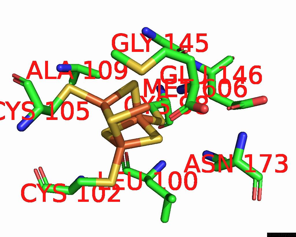
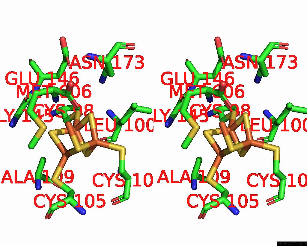
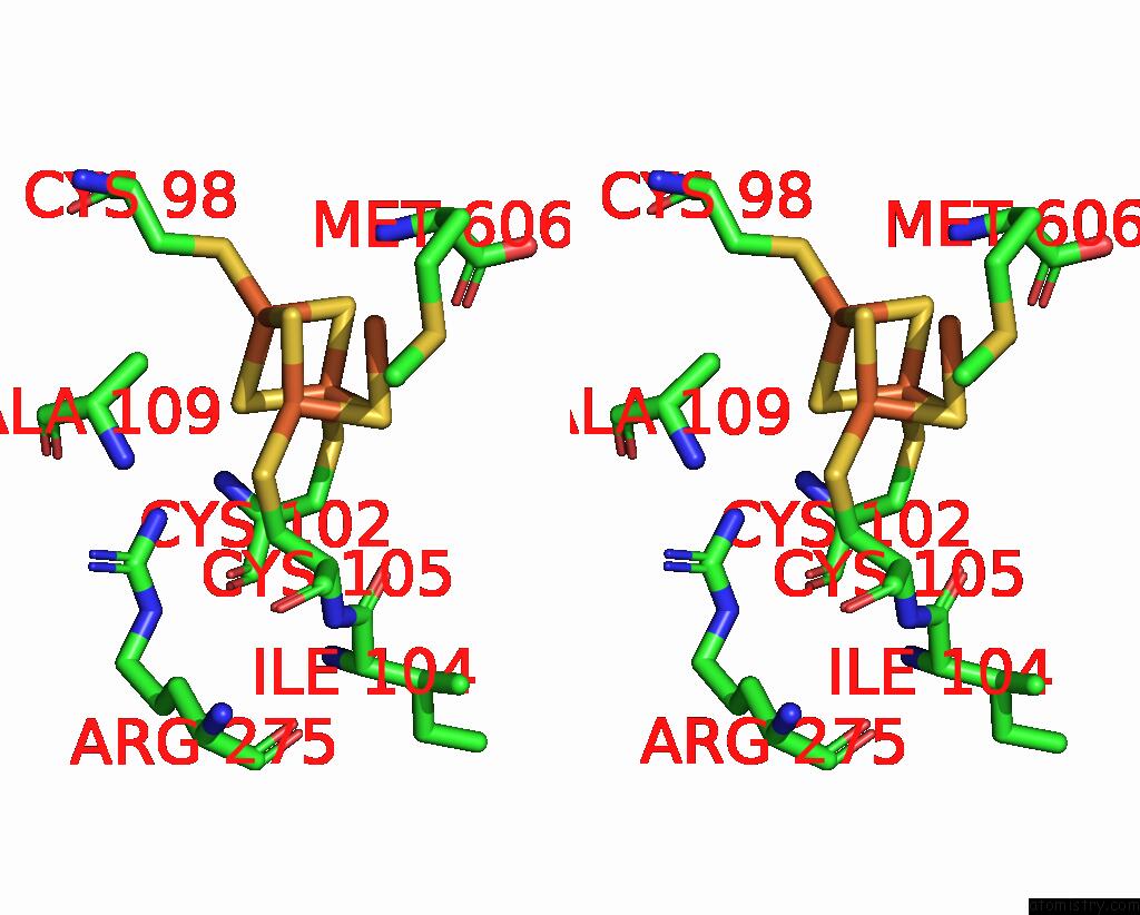
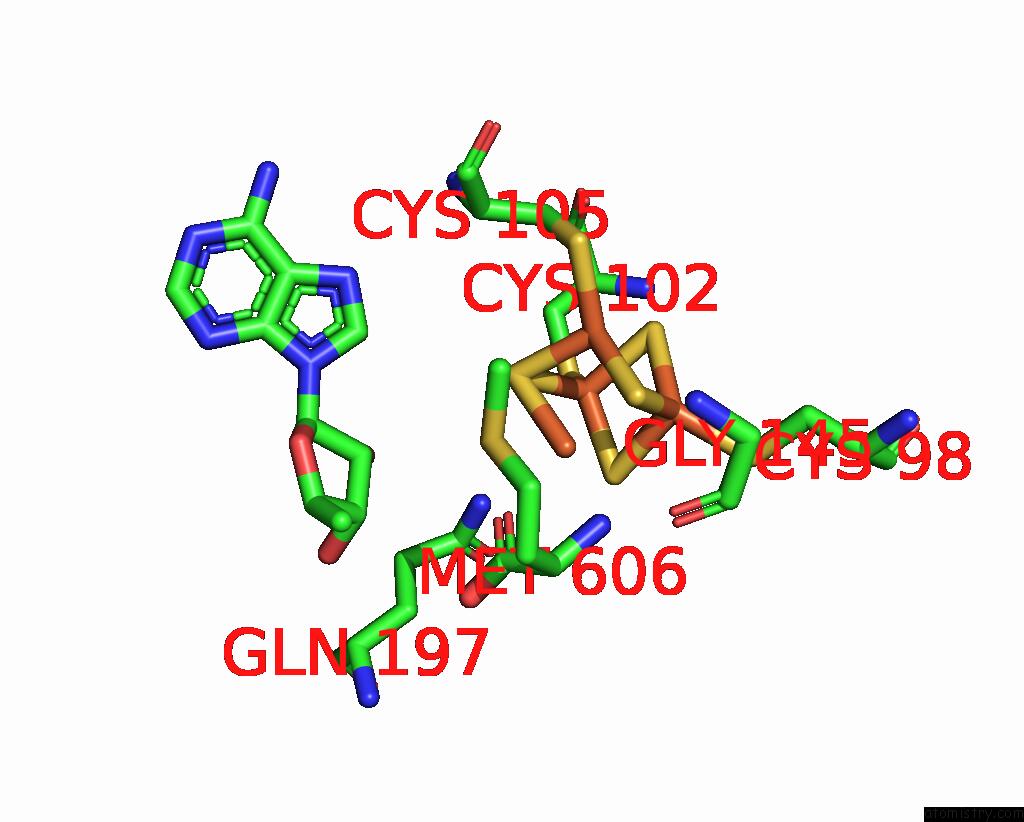
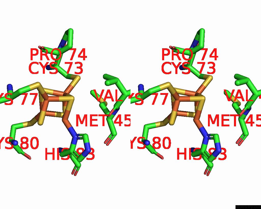
Iron binding site 1 out of 13 in 7tom
Go back to
Iron binding site 1 out
of 13 in the X-Ray Crystal Structure of Glycerol Dibiphytanyl Glycerol Tetraether - Macrocyclic Archaeol Synthase (Gdgt-Mas) From Methanocaldococcus Jannaschii with Bacterial Lipid Substrate Analog, 5'Deoxyadenosine, and Methionine Bound

Mono view

Stereo pair view
Mono view
Stereo pair view
A full contact list of Iron with other atoms in the Fe binding
site number 1 of X-Ray Crystal Structure of Glycerol Dibiphytanyl Glycerol Tetraether - Macrocyclic Archaeol Synthase (Gdgt-Mas) From Methanocaldococcus Jannaschii with Bacterial Lipid Substrate Analog, 5'Deoxyadenosine, and Methionine Bound within 5.0Å range:
|
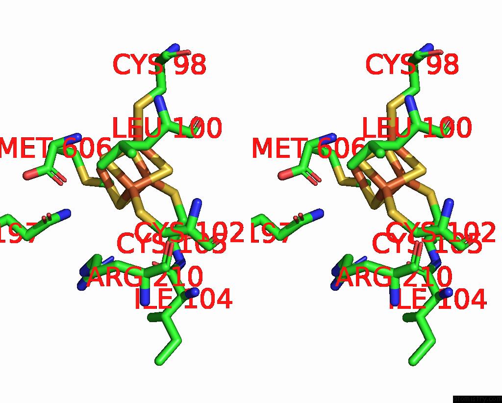
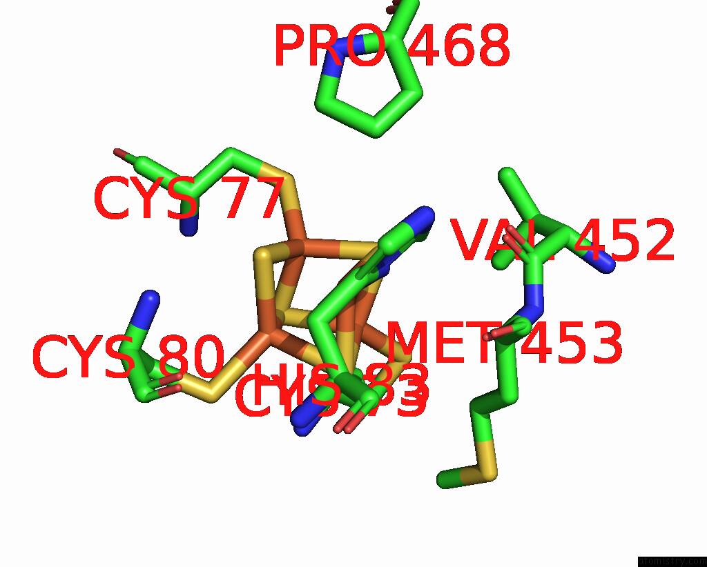
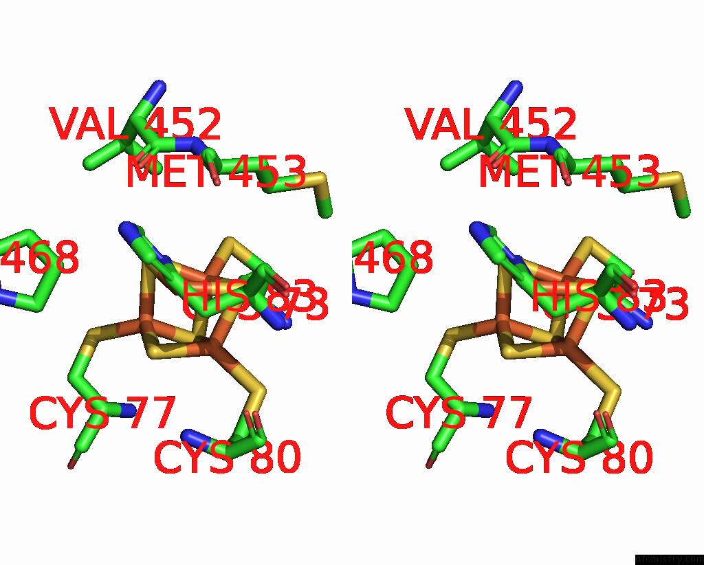
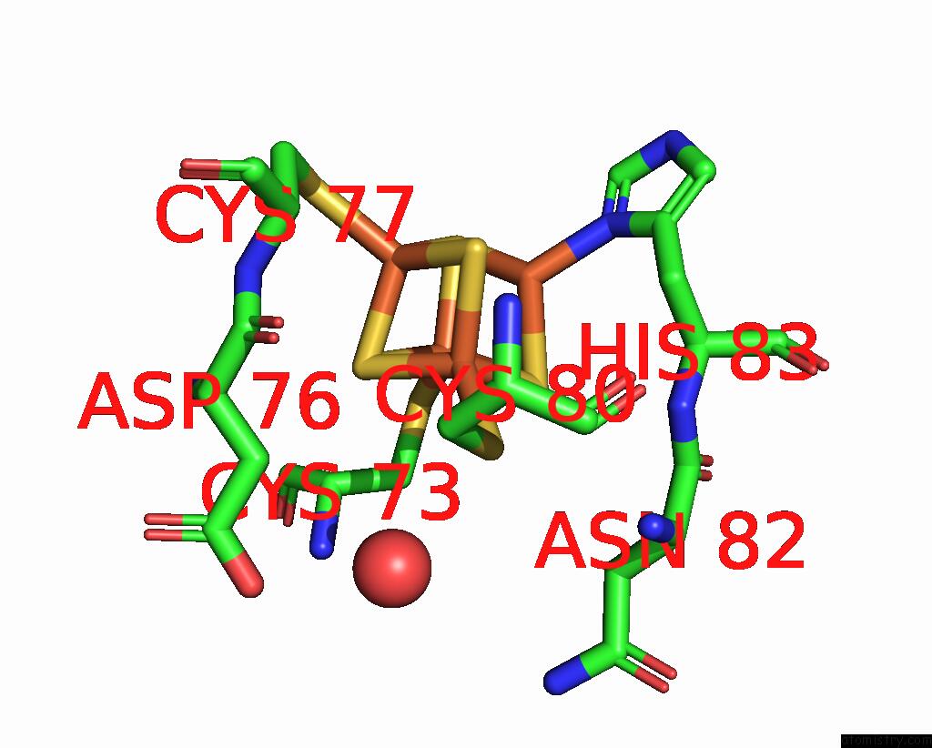
Iron binding site 2 out of 13 in 7tom
Go back to
Iron binding site 2 out
of 13 in the X-Ray Crystal Structure of Glycerol Dibiphytanyl Glycerol Tetraether - Macrocyclic Archaeol Synthase (Gdgt-Mas) From Methanocaldococcus Jannaschii with Bacterial Lipid Substrate Analog, 5'Deoxyadenosine, and Methionine Bound
Mono view
Stereo pair view
Mono view
Stereo pair view
A full contact list of Iron with other atoms in the Fe binding
site number 2 of X-Ray Crystal Structure of Glycerol Dibiphytanyl Glycerol Tetraether - Macrocyclic Archaeol Synthase (Gdgt-Mas) From Methanocaldococcus Jannaschii with Bacterial Lipid Substrate Analog, 5'Deoxyadenosine, and Methionine Bound within 5.0Å range:
|
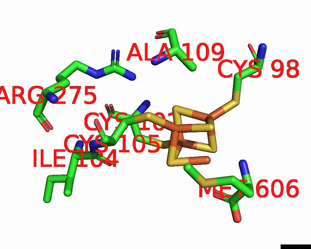
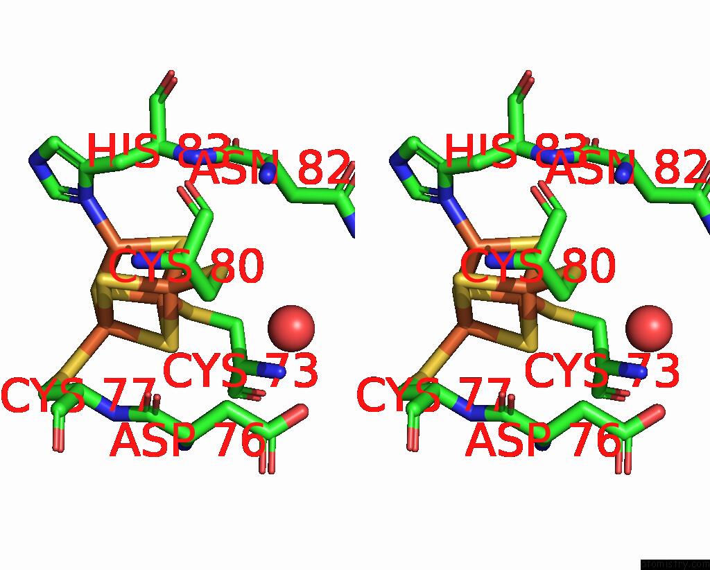
Iron binding site 3 out of 13 in 7tom
Go back to
Iron binding site 3 out
of 13 in the X-Ray Crystal Structure of Glycerol Dibiphytanyl Glycerol Tetraether - Macrocyclic Archaeol Synthase (Gdgt-Mas) From Methanocaldococcus Jannaschii with Bacterial Lipid Substrate Analog, 5'Deoxyadenosine, and Methionine Bound

Mono view
Stereo pair view
Mono view
Stereo pair view
A full contact list of Iron with other atoms in the Fe binding
site number 3 of X-Ray Crystal Structure of Glycerol Dibiphytanyl Glycerol Tetraether - Macrocyclic Archaeol Synthase (Gdgt-Mas) From Methanocaldococcus Jannaschii with Bacterial Lipid Substrate Analog, 5'Deoxyadenosine, and Methionine Bound within 5.0Å range:
|
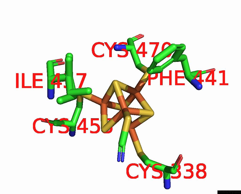
Iron binding site 4 out of 13 in 7tom
Go back to
Iron binding site 4 out
of 13 in the X-Ray Crystal Structure of Glycerol Dibiphytanyl Glycerol Tetraether - Macrocyclic Archaeol Synthase (Gdgt-Mas) From Methanocaldococcus Jannaschii with Bacterial Lipid Substrate Analog, 5'Deoxyadenosine, and Methionine Bound
Mono view
Stereo pair view
Mono view
Stereo pair view
A full contact list of Iron with other atoms in the Fe binding
site number 4 of X-Ray Crystal Structure of Glycerol Dibiphytanyl Glycerol Tetraether - Macrocyclic Archaeol Synthase (Gdgt-Mas) From Methanocaldococcus Jannaschii with Bacterial Lipid Substrate Analog, 5'Deoxyadenosine, and Methionine Bound within 5.0Å range:
|
Iron binding site 5 out of 13 in 7tom
Go back to
Iron binding site 5 out
of 13 in the X-Ray Crystal Structure of Glycerol Dibiphytanyl Glycerol Tetraether - Macrocyclic Archaeol Synthase (Gdgt-Mas) From Methanocaldococcus Jannaschii with Bacterial Lipid Substrate Analog, 5'Deoxyadenosine, and Methionine Bound
Mono view

Stereo pair view
Mono view
Stereo pair view
A full contact list of Iron with other atoms in the Fe binding
site number 5 of X-Ray Crystal Structure of Glycerol Dibiphytanyl Glycerol Tetraether - Macrocyclic Archaeol Synthase (Gdgt-Mas) From Methanocaldococcus Jannaschii with Bacterial Lipid Substrate Analog, 5'Deoxyadenosine, and Methionine Bound within 5.0Å range:
|
Iron binding site 6 out of 13 in 7tom
Go back to
Iron binding site 6 out
of 13 in the X-Ray Crystal Structure of Glycerol Dibiphytanyl Glycerol Tetraether - Macrocyclic Archaeol Synthase (Gdgt-Mas) From Methanocaldococcus Jannaschii with Bacterial Lipid Substrate Analog, 5'Deoxyadenosine, and Methionine Bound

Mono view

Stereo pair view
Mono view
Stereo pair view
A full contact list of Iron with other atoms in the Fe binding
site number 6 of X-Ray Crystal Structure of Glycerol Dibiphytanyl Glycerol Tetraether - Macrocyclic Archaeol Synthase (Gdgt-Mas) From Methanocaldococcus Jannaschii with Bacterial Lipid Substrate Analog, 5'Deoxyadenosine, and Methionine Bound within 5.0Å range:
|
Iron binding site 7 out of 13 in 7tom
Go back to
Iron binding site 7 out
of 13 in the X-Ray Crystal Structure of Glycerol Dibiphytanyl Glycerol Tetraether - Macrocyclic Archaeol Synthase (Gdgt-Mas) From Methanocaldococcus Jannaschii with Bacterial Lipid Substrate Analog, 5'Deoxyadenosine, and Methionine Bound
Mono view
Stereo pair view
Mono view
Stereo pair view
A full contact list of Iron with other atoms in the Fe binding
site number 7 of X-Ray Crystal Structure of Glycerol Dibiphytanyl Glycerol Tetraether - Macrocyclic Archaeol Synthase (Gdgt-Mas) From Methanocaldococcus Jannaschii with Bacterial Lipid Substrate Analog, 5'Deoxyadenosine, and Methionine Bound within 5.0Å range:
|
Iron binding site 8 out of 13 in 7tom
Go back to
Iron binding site 8 out
of 13 in the X-Ray Crystal Structure of Glycerol Dibiphytanyl Glycerol Tetraether - Macrocyclic Archaeol Synthase (Gdgt-Mas) From Methanocaldococcus Jannaschii with Bacterial Lipid Substrate Analog, 5'Deoxyadenosine, and Methionine Bound

Mono view
Stereo pair view
Mono view
Stereo pair view
A full contact list of Iron with other atoms in the Fe binding
site number 8 of X-Ray Crystal Structure of Glycerol Dibiphytanyl Glycerol Tetraether - Macrocyclic Archaeol Synthase (Gdgt-Mas) From Methanocaldococcus Jannaschii with Bacterial Lipid Substrate Analog, 5'Deoxyadenosine, and Methionine Bound within 5.0Å range:
|
Iron binding site 9 out of 13 in 7tom
Go back to
Iron binding site 9 out
of 13 in the X-Ray Crystal Structure of Glycerol Dibiphytanyl Glycerol Tetraether - Macrocyclic Archaeol Synthase (Gdgt-Mas) From Methanocaldococcus Jannaschii with Bacterial Lipid Substrate Analog, 5'Deoxyadenosine, and Methionine Bound
Mono view
Stereo pair view
Mono view
Stereo pair view
A full contact list of Iron with other atoms in the Fe binding
site number 9 of X-Ray Crystal Structure of Glycerol Dibiphytanyl Glycerol Tetraether - Macrocyclic Archaeol Synthase (Gdgt-Mas) From Methanocaldococcus Jannaschii with Bacterial Lipid Substrate Analog, 5'Deoxyadenosine, and Methionine Bound within 5.0Å range:
|
Iron binding site 10 out of 13 in 7tom
Go back to
Iron binding site 10 out
of 13 in the X-Ray Crystal Structure of Glycerol Dibiphytanyl Glycerol Tetraether - Macrocyclic Archaeol Synthase (Gdgt-Mas) From Methanocaldococcus Jannaschii with Bacterial Lipid Substrate Analog, 5'Deoxyadenosine, and Methionine Bound
Mono view

Stereo pair view
Mono view
Stereo pair view
A full contact list of Iron with other atoms in the Fe binding
site number 10 of X-Ray Crystal Structure of Glycerol Dibiphytanyl Glycerol Tetraether - Macrocyclic Archaeol Synthase (Gdgt-Mas) From Methanocaldococcus Jannaschii with Bacterial Lipid Substrate Analog, 5'Deoxyadenosine, and Methionine Bound within 5.0Å range:
|
Reference:
C.T.Lloyd,
D.F.Iwig,
B.Wang,
M.Cossu,
W.W.Metcalf,
A.K.Boal,
S.J.Booker.
Discovery, Structure and Mechanism of A Tetraether Lipid Synthase. Nature V. 609 197 2022.
ISSN: ESSN 1476-4687
PubMed: 35882349
DOI: 10.1038/S41586-022-05120-2
Page generated: Fri Aug 9 02:02:04 2024
ISSN: ESSN 1476-4687
PubMed: 35882349
DOI: 10.1038/S41586-022-05120-2
Last articles
Zn in 9MJ5Zn in 9HNW
Zn in 9G0L
Zn in 9FNE
Zn in 9DZN
Zn in 9E0I
Zn in 9D32
Zn in 9DAK
Zn in 8ZXC
Zn in 8ZUF