Iron »
PDB 7ttq-7us7 »
7txy »
Iron in PDB 7txy: Crystal Structure of the 2-Aminophenol 1,6-Dioxygenase From the Aro Bacterial Microcompartment of Micromonospora Rosaria
Protein crystallography data
The structure of Crystal Structure of the 2-Aminophenol 1,6-Dioxygenase From the Aro Bacterial Microcompartment of Micromonospora Rosaria, PDB code: 7txy
was solved by
M.Sutter,
L.Doron,
C.A.Kerfeld,
with X-Ray Crystallography technique. A brief refinement statistics is given in the table below:
| Resolution Low / High (Å) | 46.14 / 1.75 |
| Space group | P 1 |
| Cell size a, b, c (Å), α, β, γ (°) | 68.8, 83.855, 110.272, 85.59, 73.1, 89.26 |
| R / Rfree (%) | 19.4 / 22.2 |
Iron Binding Sites:
The binding sites of Iron atom in the Crystal Structure of the 2-Aminophenol 1,6-Dioxygenase From the Aro Bacterial Microcompartment of Micromonospora Rosaria
(pdb code 7txy). This binding sites where shown within
5.0 Angstroms radius around Iron atom.
In total 4 binding sites of Iron where determined in the Crystal Structure of the 2-Aminophenol 1,6-Dioxygenase From the Aro Bacterial Microcompartment of Micromonospora Rosaria, PDB code: 7txy:
Jump to Iron binding site number: 1; 2; 3; 4;
In total 4 binding sites of Iron where determined in the Crystal Structure of the 2-Aminophenol 1,6-Dioxygenase From the Aro Bacterial Microcompartment of Micromonospora Rosaria, PDB code: 7txy:
Jump to Iron binding site number: 1; 2; 3; 4;
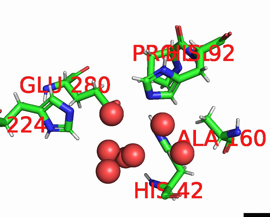
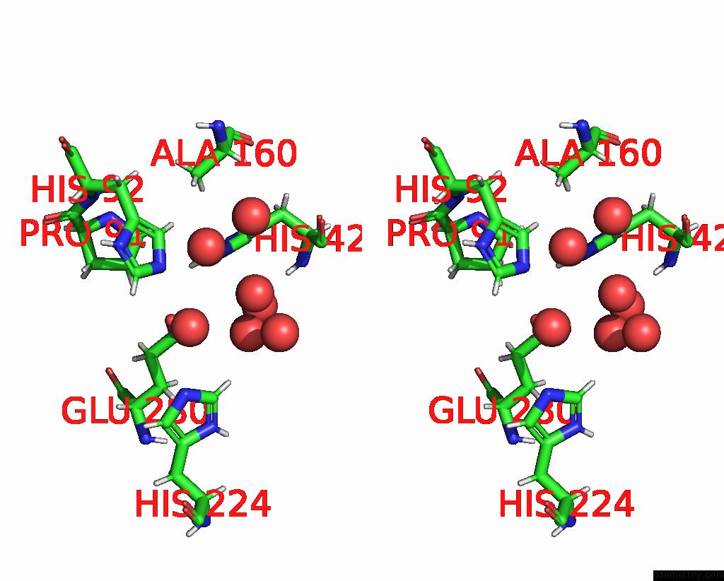
Iron binding site 1 out of 4 in 7txy
Go back to
Iron binding site 1 out
of 4 in the Crystal Structure of the 2-Aminophenol 1,6-Dioxygenase From the Aro Bacterial Microcompartment of Micromonospora Rosaria
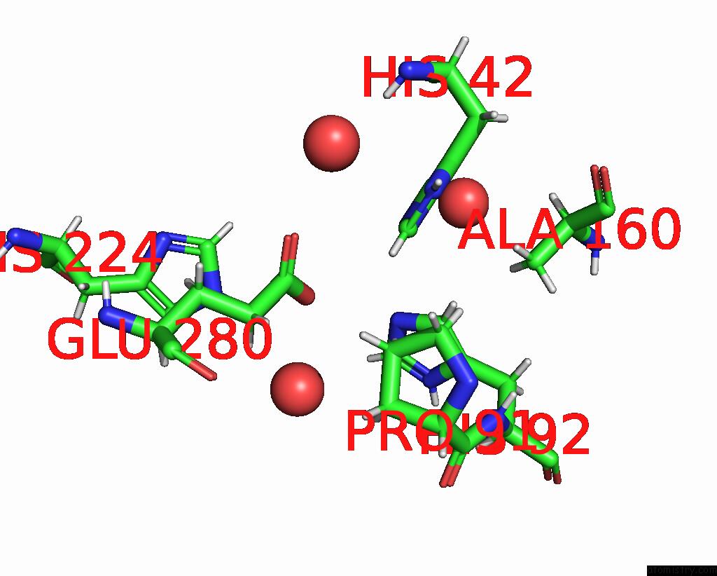
Mono view
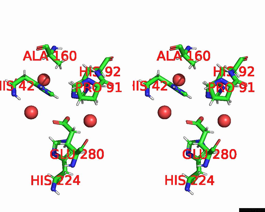
Stereo pair view
Mono view
Stereo pair view
A full contact list of Iron with other atoms in the Fe binding
site number 1 of Crystal Structure of the 2-Aminophenol 1,6-Dioxygenase From the Aro Bacterial Microcompartment of Micromonospora Rosaria within 5.0Å range:
|
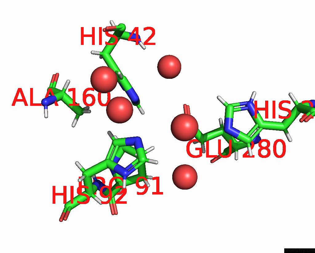
Iron binding site 2 out of 4 in 7txy
Go back to
Iron binding site 2 out
of 4 in the Crystal Structure of the 2-Aminophenol 1,6-Dioxygenase From the Aro Bacterial Microcompartment of Micromonospora Rosaria
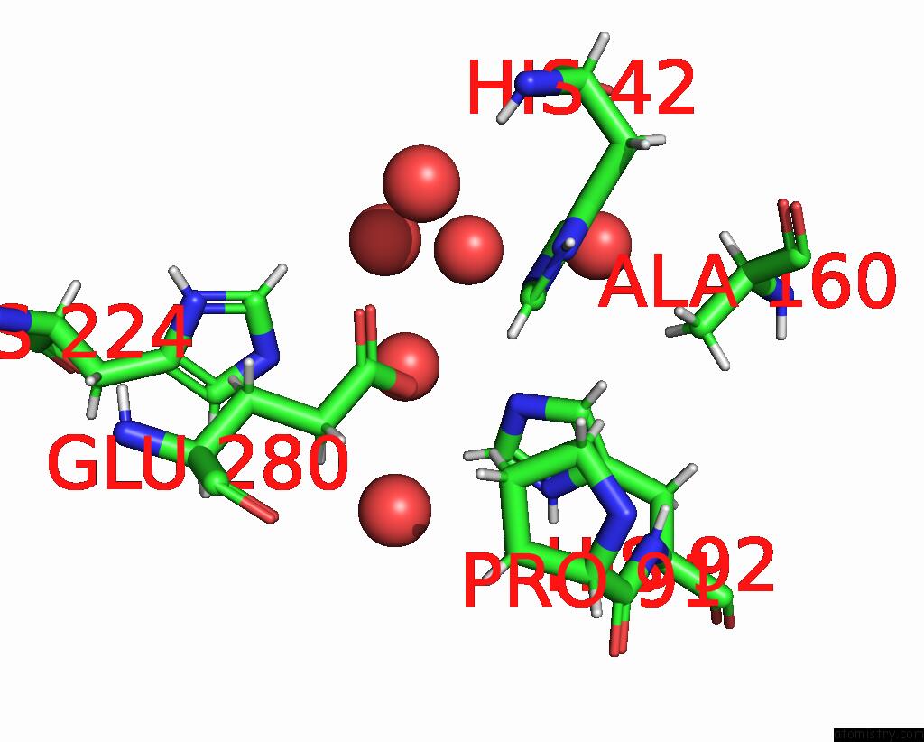
Mono view
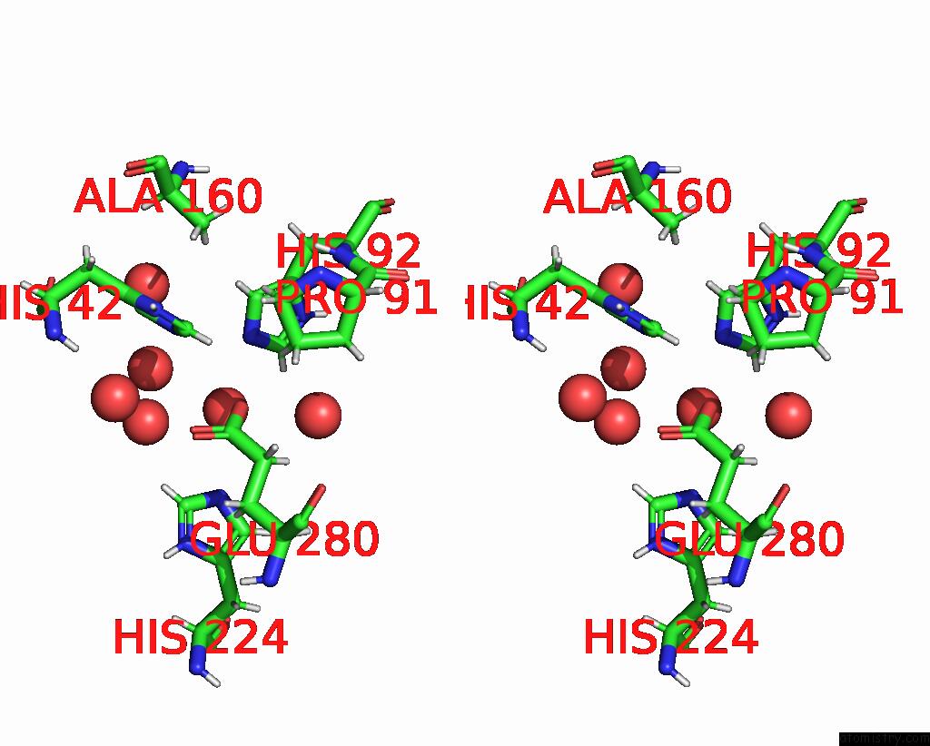
Stereo pair view
Mono view
Stereo pair view
A full contact list of Iron with other atoms in the Fe binding
site number 2 of Crystal Structure of the 2-Aminophenol 1,6-Dioxygenase From the Aro Bacterial Microcompartment of Micromonospora Rosaria within 5.0Å range:
|
Iron binding site 3 out of 4 in 7txy
Go back to
Iron binding site 3 out
of 4 in the Crystal Structure of the 2-Aminophenol 1,6-Dioxygenase From the Aro Bacterial Microcompartment of Micromonospora Rosaria
Mono view
Stereo pair view
Mono view
Stereo pair view
A full contact list of Iron with other atoms in the Fe binding
site number 3 of Crystal Structure of the 2-Aminophenol 1,6-Dioxygenase From the Aro Bacterial Microcompartment of Micromonospora Rosaria within 5.0Å range:
|
Iron binding site 4 out of 4 in 7txy
Go back to
Iron binding site 4 out
of 4 in the Crystal Structure of the 2-Aminophenol 1,6-Dioxygenase From the Aro Bacterial Microcompartment of Micromonospora Rosaria
Mono view
Stereo pair view
Mono view
Stereo pair view
A full contact list of Iron with other atoms in the Fe binding
site number 4 of Crystal Structure of the 2-Aminophenol 1,6-Dioxygenase From the Aro Bacterial Microcompartment of Micromonospora Rosaria within 5.0Å range:
|
Reference:
L.Doron,
M.Sutter,
C.A.Kerfeld.
Characterization of A Novel Aromatic Substrate Processing Microcompartment in Actinobacteria To Be Published.
Page generated: Fri Aug 9 02:40:51 2024
Last articles
F in 7RE0F in 7RDY
F in 7RDX
F in 7RD7
F in 7RD6
F in 7RA5
F in 7RBT
F in 7R9Y
F in 7R9N
F in 7R9Z