Iron »
PDB 7usn-7v43 »
7uur »
Iron in PDB 7uur: The 1.67 Angstrom Cryoem Structure of the [Nife]-Hydrogenase Huc From Mycobacterium Smegmatis - Catalytic Dimer (HUC2S2L)
Enzymatic activity of The 1.67 Angstrom Cryoem Structure of the [Nife]-Hydrogenase Huc From Mycobacterium Smegmatis - Catalytic Dimer (HUC2S2L)
All present enzymatic activity of The 1.67 Angstrom Cryoem Structure of the [Nife]-Hydrogenase Huc From Mycobacterium Smegmatis - Catalytic Dimer (HUC2S2L):
1.12.99.6;
1.12.99.6;
Other elements in 7uur:
The structure of The 1.67 Angstrom Cryoem Structure of the [Nife]-Hydrogenase Huc From Mycobacterium Smegmatis - Catalytic Dimer (HUC2S2L) also contains other interesting chemical elements:
| Nickel | (Ni) | 2 atoms |
| Magnesium | (Mg) | 2 atoms |
Iron Binding Sites:
Pages:
>>> Page 1 <<< Page 2, Binding sites: 11 - 20;Binding sites:
The binding sites of Iron atom in the The 1.67 Angstrom Cryoem Structure of the [Nife]-Hydrogenase Huc From Mycobacterium Smegmatis - Catalytic Dimer (HUC2S2L) (pdb code 7uur). This binding sites where shown within 5.0 Angstroms radius around Iron atom.In total 20 binding sites of Iron where determined in the The 1.67 Angstrom Cryoem Structure of the [Nife]-Hydrogenase Huc From Mycobacterium Smegmatis - Catalytic Dimer (HUC2S2L), PDB code: 7uur:
Jump to Iron binding site number: 1; 2; 3; 4; 5; 6; 7; 8; 9; 10;
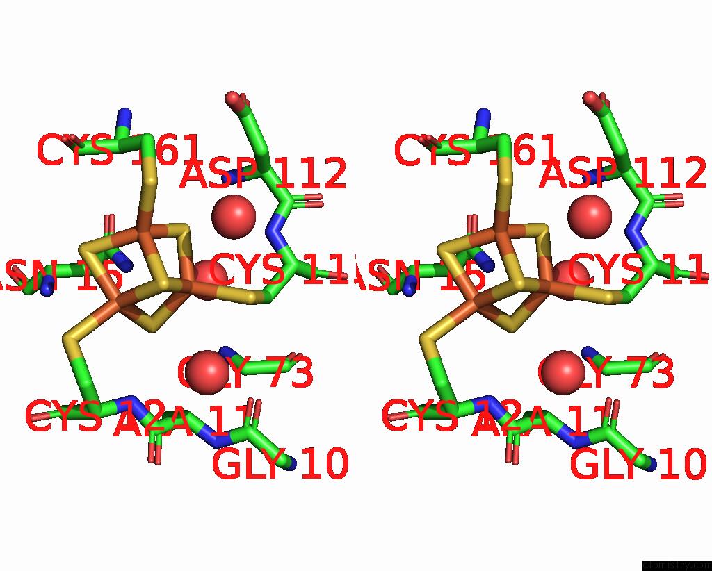
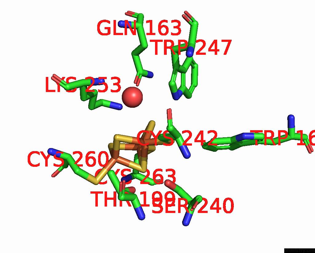
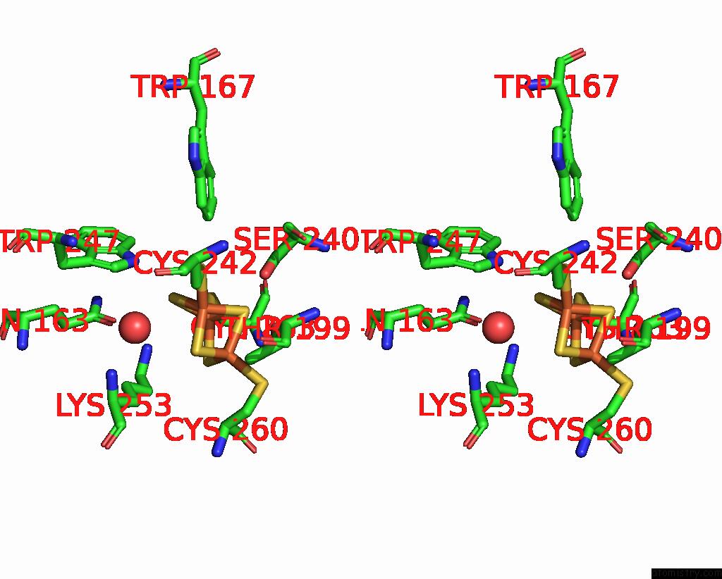
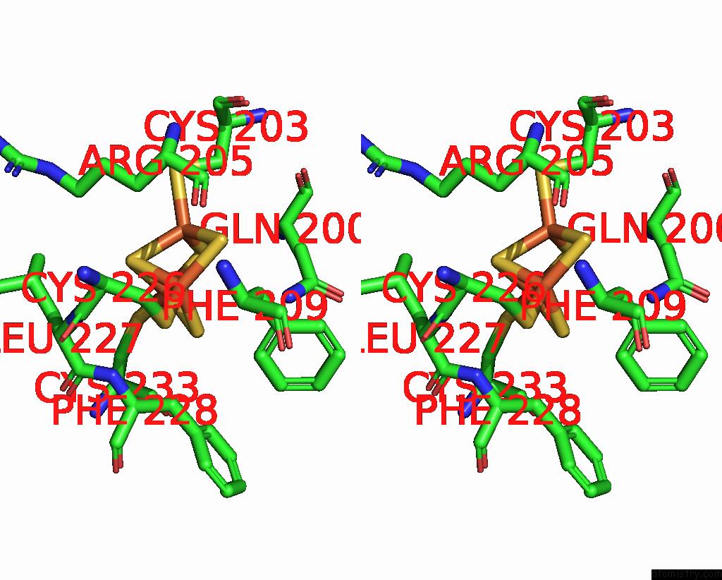
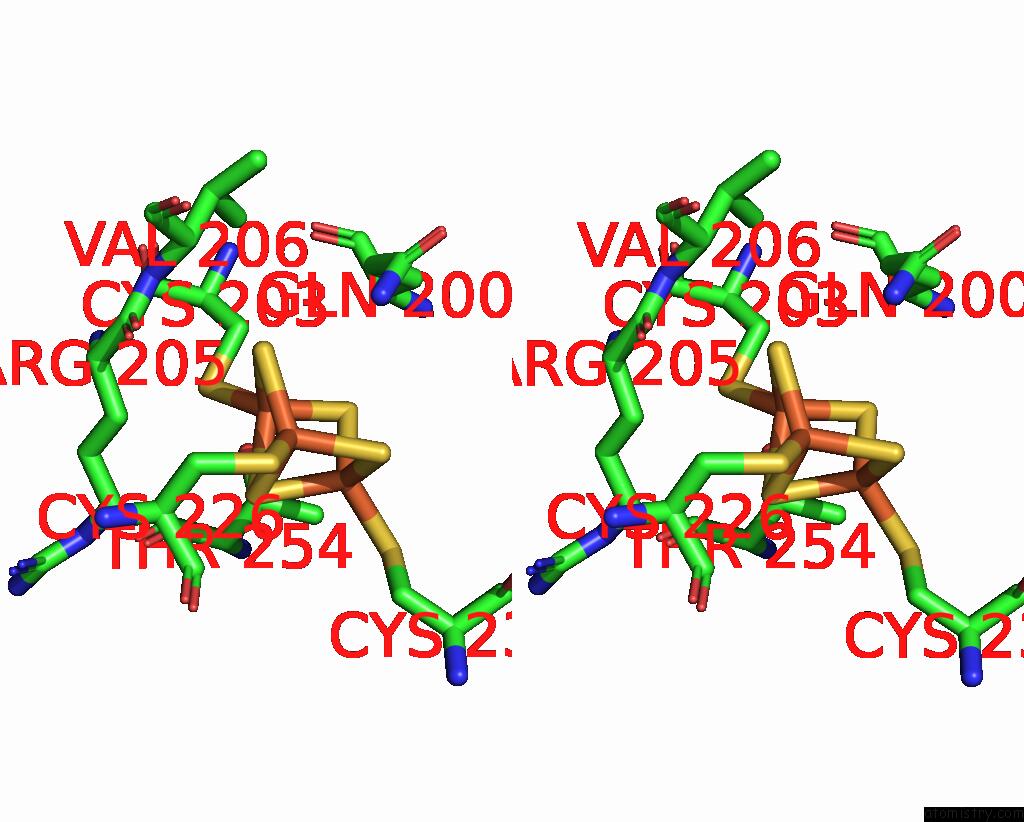
Iron binding site 1 out of 20 in 7uur
Go back to
Iron binding site 1 out
of 20 in the The 1.67 Angstrom Cryoem Structure of the [Nife]-Hydrogenase Huc From Mycobacterium Smegmatis - Catalytic Dimer (HUC2S2L)
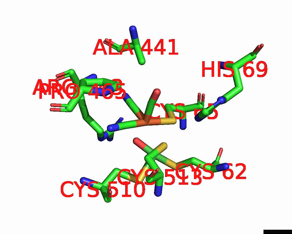
Mono view
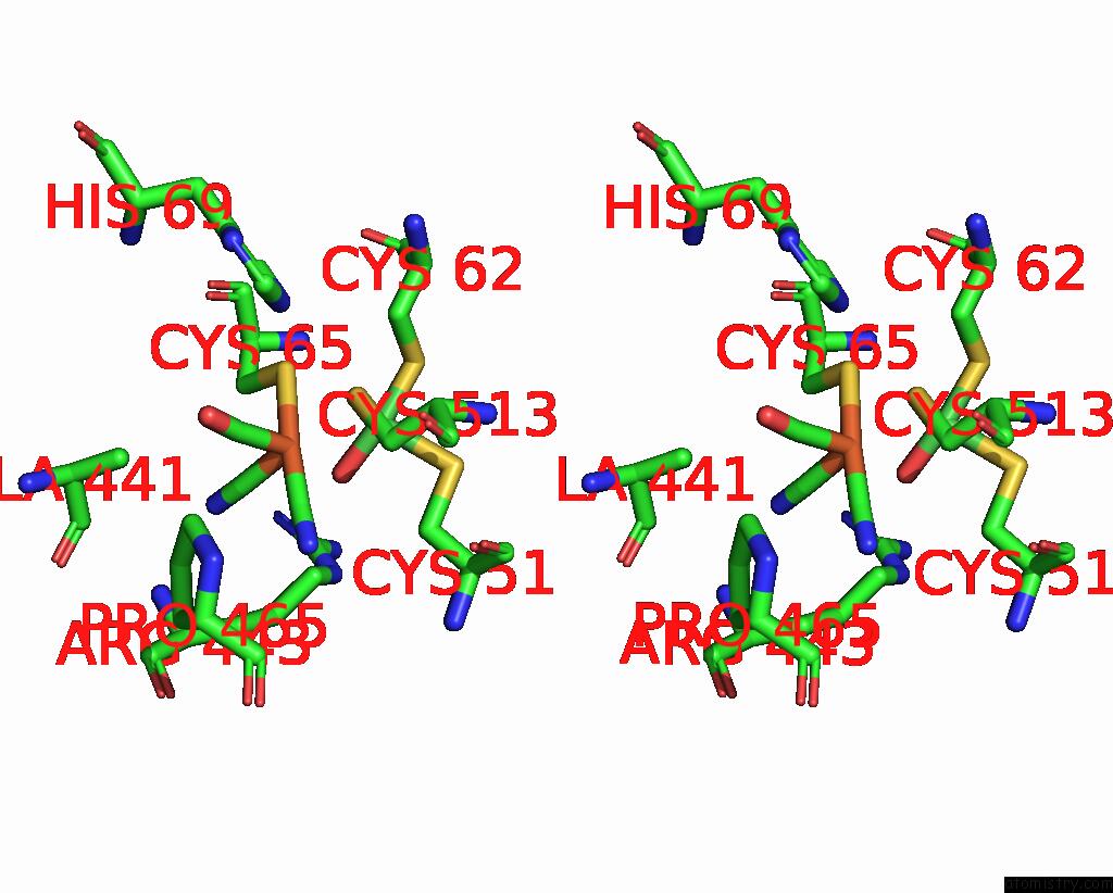
Stereo pair view
Mono view
Stereo pair view
A full contact list of Iron with other atoms in the Fe binding
site number 1 of The 1.67 Angstrom Cryoem Structure of the [Nife]-Hydrogenase Huc From Mycobacterium Smegmatis - Catalytic Dimer (HUC2S2L) within 5.0Å range:
|
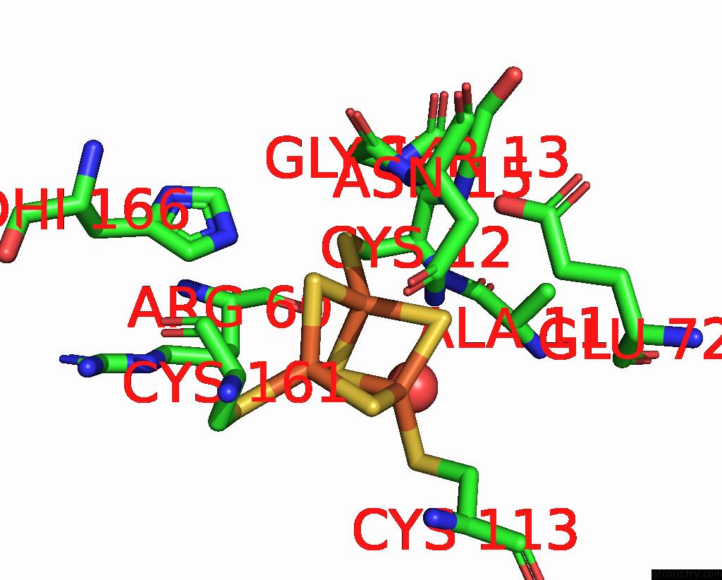
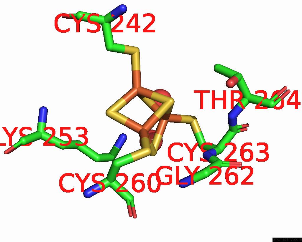
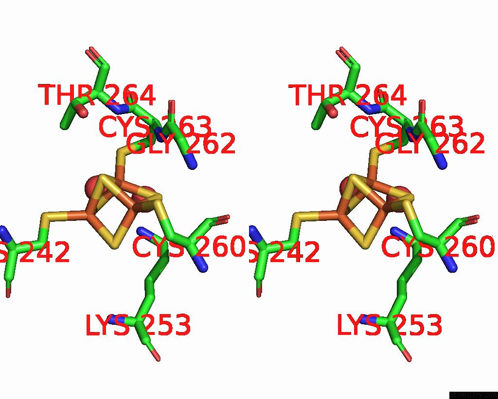
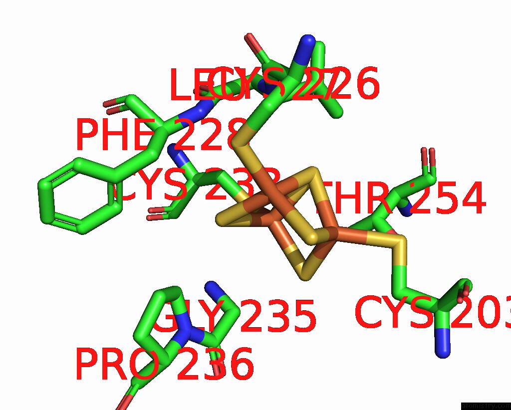
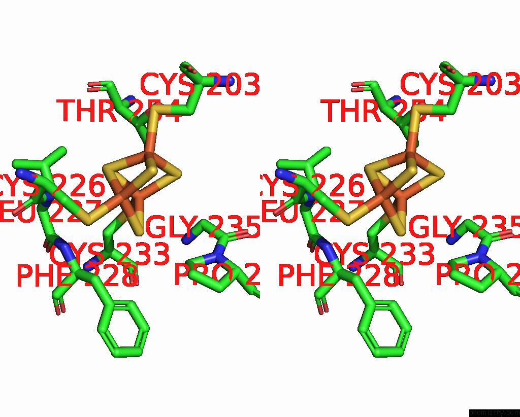
Iron binding site 2 out of 20 in 7uur
Go back to
Iron binding site 2 out
of 20 in the The 1.67 Angstrom Cryoem Structure of the [Nife]-Hydrogenase Huc From Mycobacterium Smegmatis - Catalytic Dimer (HUC2S2L)
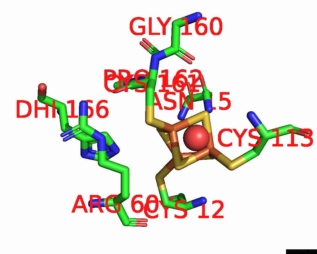
Mono view
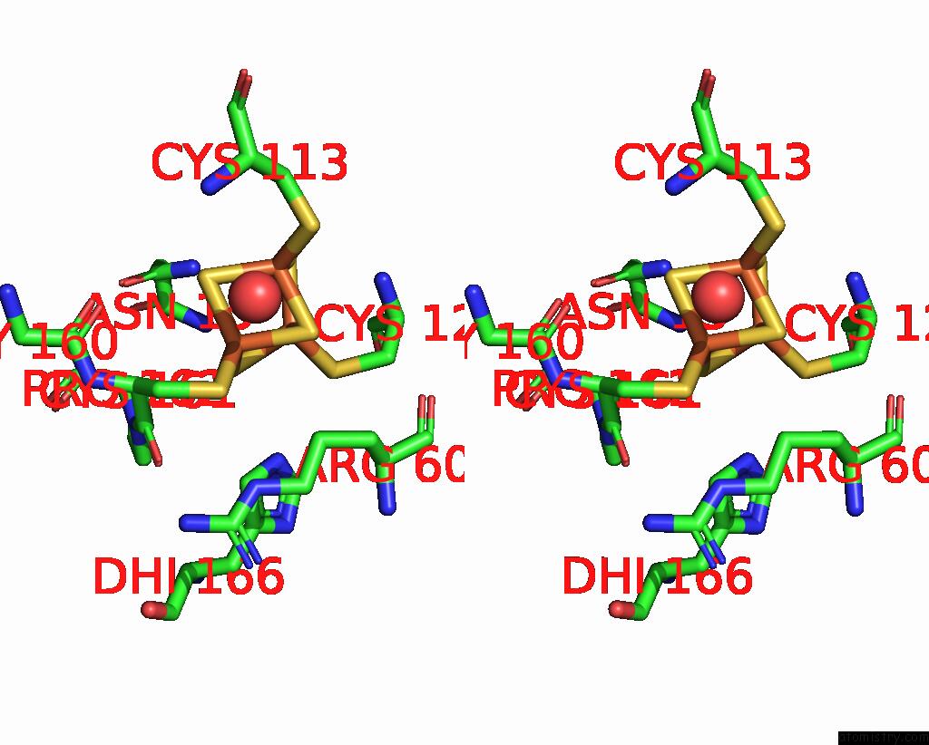
Stereo pair view
Mono view
Stereo pair view
A full contact list of Iron with other atoms in the Fe binding
site number 2 of The 1.67 Angstrom Cryoem Structure of the [Nife]-Hydrogenase Huc From Mycobacterium Smegmatis - Catalytic Dimer (HUC2S2L) within 5.0Å range:
|
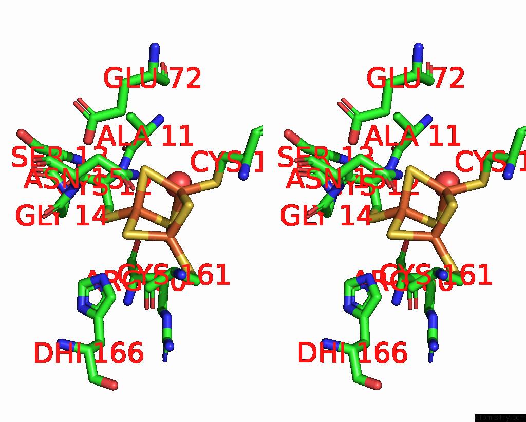
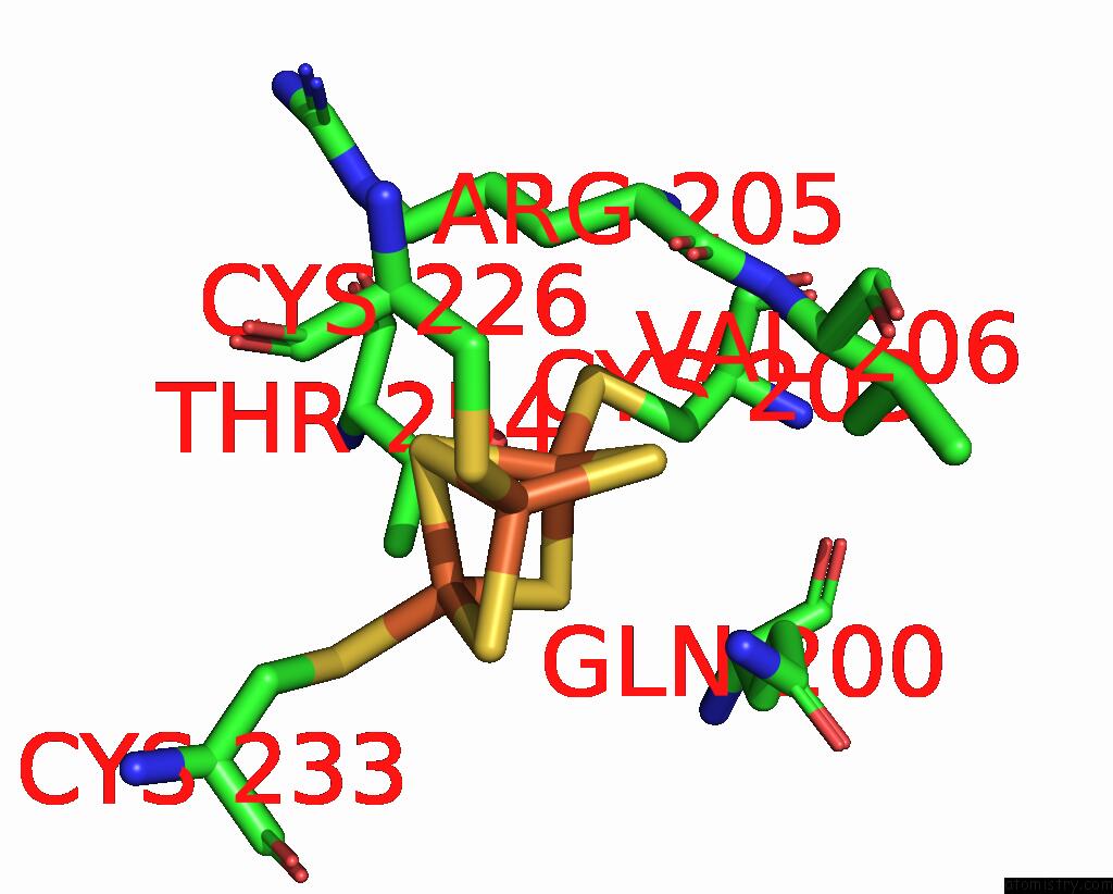
Iron binding site 3 out of 20 in 7uur
Go back to
Iron binding site 3 out
of 20 in the The 1.67 Angstrom Cryoem Structure of the [Nife]-Hydrogenase Huc From Mycobacterium Smegmatis - Catalytic Dimer (HUC2S2L)

Mono view
Stereo pair view
Mono view
Stereo pair view
A full contact list of Iron with other atoms in the Fe binding
site number 3 of The 1.67 Angstrom Cryoem Structure of the [Nife]-Hydrogenase Huc From Mycobacterium Smegmatis - Catalytic Dimer (HUC2S2L) within 5.0Å range:
|
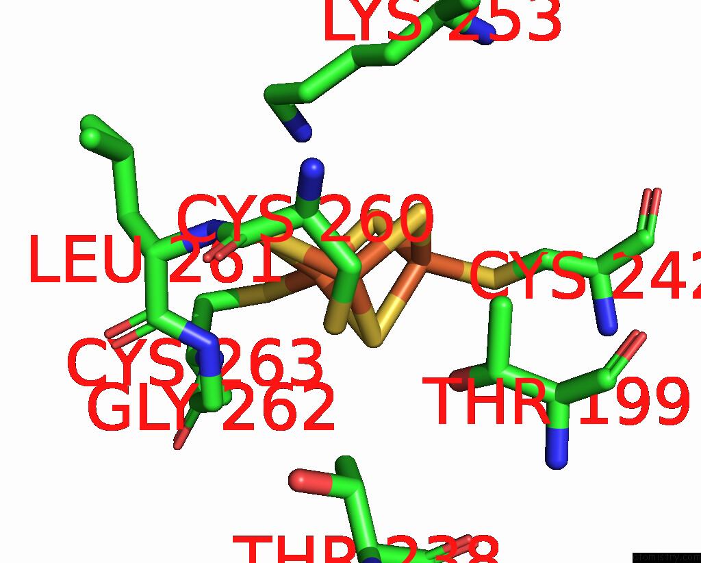
Iron binding site 4 out of 20 in 7uur
Go back to
Iron binding site 4 out
of 20 in the The 1.67 Angstrom Cryoem Structure of the [Nife]-Hydrogenase Huc From Mycobacterium Smegmatis - Catalytic Dimer (HUC2S2L)
Mono view
Stereo pair view
Mono view
Stereo pair view
A full contact list of Iron with other atoms in the Fe binding
site number 4 of The 1.67 Angstrom Cryoem Structure of the [Nife]-Hydrogenase Huc From Mycobacterium Smegmatis - Catalytic Dimer (HUC2S2L) within 5.0Å range:
|
Iron binding site 5 out of 20 in 7uur
Go back to
Iron binding site 5 out
of 20 in the The 1.67 Angstrom Cryoem Structure of the [Nife]-Hydrogenase Huc From Mycobacterium Smegmatis - Catalytic Dimer (HUC2S2L)
Mono view

Stereo pair view
Mono view
Stereo pair view
A full contact list of Iron with other atoms in the Fe binding
site number 5 of The 1.67 Angstrom Cryoem Structure of the [Nife]-Hydrogenase Huc From Mycobacterium Smegmatis - Catalytic Dimer (HUC2S2L) within 5.0Å range:
|
Iron binding site 6 out of 20 in 7uur
Go back to
Iron binding site 6 out
of 20 in the The 1.67 Angstrom Cryoem Structure of the [Nife]-Hydrogenase Huc From Mycobacterium Smegmatis - Catalytic Dimer (HUC2S2L)
Mono view
Stereo pair view
Mono view
Stereo pair view
A full contact list of Iron with other atoms in the Fe binding
site number 6 of The 1.67 Angstrom Cryoem Structure of the [Nife]-Hydrogenase Huc From Mycobacterium Smegmatis - Catalytic Dimer (HUC2S2L) within 5.0Å range:
|
Iron binding site 7 out of 20 in 7uur
Go back to
Iron binding site 7 out
of 20 in the The 1.67 Angstrom Cryoem Structure of the [Nife]-Hydrogenase Huc From Mycobacterium Smegmatis - Catalytic Dimer (HUC2S2L)
Mono view
Stereo pair view
Mono view
Stereo pair view
A full contact list of Iron with other atoms in the Fe binding
site number 7 of The 1.67 Angstrom Cryoem Structure of the [Nife]-Hydrogenase Huc From Mycobacterium Smegmatis - Catalytic Dimer (HUC2S2L) within 5.0Å range:
|
Iron binding site 8 out of 20 in 7uur
Go back to
Iron binding site 8 out
of 20 in the The 1.67 Angstrom Cryoem Structure of the [Nife]-Hydrogenase Huc From Mycobacterium Smegmatis - Catalytic Dimer (HUC2S2L)

Mono view
Stereo pair view
Mono view
Stereo pair view
A full contact list of Iron with other atoms in the Fe binding
site number 8 of The 1.67 Angstrom Cryoem Structure of the [Nife]-Hydrogenase Huc From Mycobacterium Smegmatis - Catalytic Dimer (HUC2S2L) within 5.0Å range:
|
Iron binding site 9 out of 20 in 7uur
Go back to
Iron binding site 9 out
of 20 in the The 1.67 Angstrom Cryoem Structure of the [Nife]-Hydrogenase Huc From Mycobacterium Smegmatis - Catalytic Dimer (HUC2S2L)
Mono view
Stereo pair view
Mono view
Stereo pair view
A full contact list of Iron with other atoms in the Fe binding
site number 9 of The 1.67 Angstrom Cryoem Structure of the [Nife]-Hydrogenase Huc From Mycobacterium Smegmatis - Catalytic Dimer (HUC2S2L) within 5.0Å range:
|
Iron binding site 10 out of 20 in 7uur
Go back to
Iron binding site 10 out
of 20 in the The 1.67 Angstrom Cryoem Structure of the [Nife]-Hydrogenase Huc From Mycobacterium Smegmatis - Catalytic Dimer (HUC2S2L)
Mono view
Stereo pair view
Mono view
Stereo pair view
A full contact list of Iron with other atoms in the Fe binding
site number 10 of The 1.67 Angstrom Cryoem Structure of the [Nife]-Hydrogenase Huc From Mycobacterium Smegmatis - Catalytic Dimer (HUC2S2L) within 5.0Å range:
|
Reference:
R.Grinter,
A.Kropp,
H.Venugopal,
M.Senger,
J.Badley,
P.Cabotaje,
S.T.Stripp,
C.K.Barlow,
M.Belousoff,
G.M.Cook,
R.B.Schittenhelm,
S.Khalid,
G.Berggren,
G.Greening.
An Oxygen-Insensitive, Quinone-Transporting Hydrogenase Enables Bacteria to Extract Energy From Air To Be Published.
Page generated: Thu Aug 7 06:36:54 2025
Last articles
Fe in 7W35Fe in 7W3E
Fe in 7W32
Fe in 7W2Y
Fe in 7W31
Fe in 7W2U
Fe in 7W2R
Fe in 7W2L
Fe in 7W2K
Fe in 7W2J