Iron »
PDB 7v43-7voy »
7vde »
Iron in PDB 7vde: 3.6 A Structure of the Human Hemoglobin
Iron Binding Sites:
The binding sites of Iron atom in the 3.6 A Structure of the Human Hemoglobin
(pdb code 7vde). This binding sites where shown within
5.0 Angstroms radius around Iron atom.
In total 4 binding sites of Iron where determined in the 3.6 A Structure of the Human Hemoglobin, PDB code: 7vde:
Jump to Iron binding site number: 1; 2; 3; 4;
In total 4 binding sites of Iron where determined in the 3.6 A Structure of the Human Hemoglobin, PDB code: 7vde:
Jump to Iron binding site number: 1; 2; 3; 4;
Iron binding site 1 out of 4 in 7vde
Go back to
Iron binding site 1 out
of 4 in the 3.6 A Structure of the Human Hemoglobin

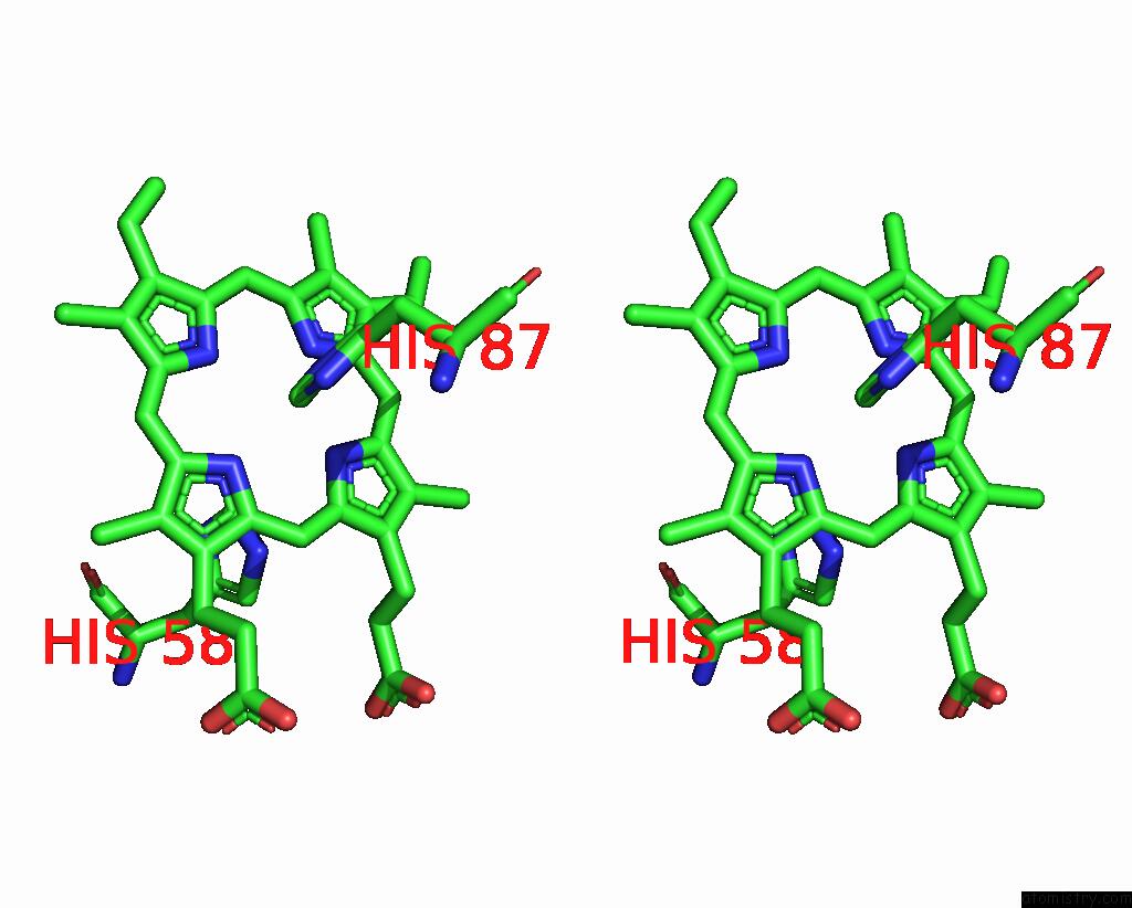
Mono view

Stereo pair view
Mono view
Stereo pair view
A full contact list of Iron with other atoms in the Fe binding
site number 1 of 3.6 A Structure of the Human Hemoglobin within 5.0Å range:
|
Iron binding site 2 out of 4 in 7vde
Go back to
Iron binding site 2 out
of 4 in the 3.6 A Structure of the Human Hemoglobin

Mono view

Stereo pair view
Mono view
Stereo pair view
A full contact list of Iron with other atoms in the Fe binding
site number 2 of 3.6 A Structure of the Human Hemoglobin within 5.0Å range:
|
Iron binding site 3 out of 4 in 7vde
Go back to
Iron binding site 3 out
of 4 in the 3.6 A Structure of the Human Hemoglobin

Mono view
Stereo pair view
Mono view
Stereo pair view
A full contact list of Iron with other atoms in the Fe binding
site number 3 of 3.6 A Structure of the Human Hemoglobin within 5.0Å range:
|
Iron binding site 4 out of 4 in 7vde
Go back to
Iron binding site 4 out
of 4 in the 3.6 A Structure of the Human Hemoglobin

Mono view

Stereo pair view
Mono view
Stereo pair view
A full contact list of Iron with other atoms in the Fe binding
site number 4 of 3.6 A Structure of the Human Hemoglobin within 5.0Å range:
|
Reference:
H.Fan,
B.Wang,
Y.Zhang,
Y.Zhu,
B.Song,
H.Xu,
Y.Zhai,
M.Qiao,
F.Sun.
A Cryo-Electron Microscopy Support Film Formed By 2D Crystals of Hydrophobin Hfbi. Nat Commun V. 12 7257 2021.
ISSN: ESSN 2041-1723
PubMed: 34907237
DOI: 10.1038/S41467-021-27596-8
Page generated: Fri Aug 9 05:28:18 2024
ISSN: ESSN 2041-1723
PubMed: 34907237
DOI: 10.1038/S41467-021-27596-8
Last articles
Zn in 9MJ5Zn in 9HNW
Zn in 9G0L
Zn in 9FNE
Zn in 9DZN
Zn in 9E0I
Zn in 9D32
Zn in 9DAK
Zn in 8ZXC
Zn in 8ZUF