Iron »
PDB 7v43-7voy »
7vgm »
Iron in PDB 7vgm: Crystal Structure of Phenylalanine Hydroxylase From Bacillus Cereus Atcc 14579
Enzymatic activity of Crystal Structure of Phenylalanine Hydroxylase From Bacillus Cereus Atcc 14579
All present enzymatic activity of Crystal Structure of Phenylalanine Hydroxylase From Bacillus Cereus Atcc 14579:
1.14.16.1;
1.14.16.1;
Protein crystallography data
The structure of Crystal Structure of Phenylalanine Hydroxylase From Bacillus Cereus Atcc 14579, PDB code: 7vgm
was solved by
J.Park,
K.-J.Kim,
with X-Ray Crystallography technique. A brief refinement statistics is given in the table below:
| Resolution Low / High (Å) | 33.49 / 2.27 |
| Space group | P 21 21 21 |
| Cell size a, b, c (Å), α, β, γ (°) | 77.273, 93.698, 95.676, 90, 90, 90 |
| R / Rfree (%) | 17.3 / 24.1 |
Iron Binding Sites:
The binding sites of Iron atom in the Crystal Structure of Phenylalanine Hydroxylase From Bacillus Cereus Atcc 14579
(pdb code 7vgm). This binding sites where shown within
5.0 Angstroms radius around Iron atom.
In total only one binding site of Iron was determined in the Crystal Structure of Phenylalanine Hydroxylase From Bacillus Cereus Atcc 14579, PDB code: 7vgm:
In total only one binding site of Iron was determined in the Crystal Structure of Phenylalanine Hydroxylase From Bacillus Cereus Atcc 14579, PDB code: 7vgm:
Iron binding site 1 out of 1 in 7vgm
Go back to
Iron binding site 1 out
of 1 in the Crystal Structure of Phenylalanine Hydroxylase From Bacillus Cereus Atcc 14579
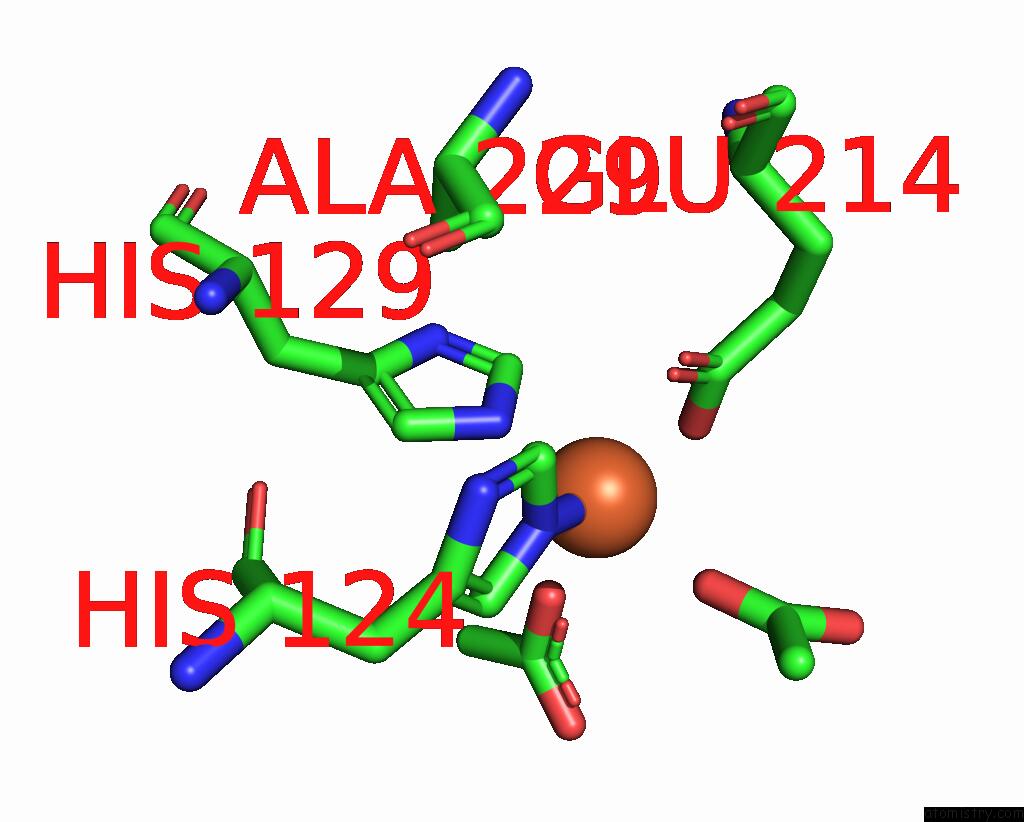
Mono view
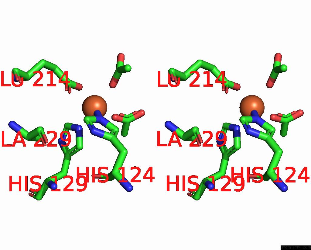
Stereo pair view
Mono view
Stereo pair view
A full contact list of Iron with other atoms in the Fe binding
site number 1 of Crystal Structure of Phenylalanine Hydroxylase From Bacillus Cereus Atcc 14579 within 5.0Å range:
|
Reference:
J.Park,
J.Hong,
J.Seok,
H.Hong,
H.Seo,
K.J.Kim.
Structural Studies of A Novel Auxiliary-Domain-Containing Phenylalanine Hydroxylase From Bacillus Cereus Atcc 14579. Acta Crystallogr D Struct V. 78 586 2022BIOL.
ISSN: ISSN 2059-7983
PubMed: 35503207
DOI: 10.1107/S2059798322002674
Page generated: Fri Aug 9 05:30:08 2024
ISSN: ISSN 2059-7983
PubMed: 35503207
DOI: 10.1107/S2059798322002674
Last articles
Zn in 9MJ5Zn in 9HNW
Zn in 9G0L
Zn in 9FNE
Zn in 9DZN
Zn in 9E0I
Zn in 9D32
Zn in 9DAK
Zn in 8ZXC
Zn in 8ZUF