Iron »
PDB 7v43-7voy »
7vos »
Iron in PDB 7vos: High-Resolution Neutron and X-Ray Joint Refined Structure of High- Potential Iron-Sulfur Protein in the Oxidized State
Protein crystallography data
The structure of High-Resolution Neutron and X-Ray Joint Refined Structure of High- Potential Iron-Sulfur Protein in the Oxidized State, PDB code: 7vos
was solved by
Y.Hanazono,
Y.Hirano,
K.Takeda,
K.Kusaka,
T.Tamada,
K.Miki,
with X-Ray Crystallography technique. A brief refinement statistics is given in the table below:
| Resolution Low / High (Å) | N/A / 0.66 |
| Space group | P 21 21 21 |
| Cell size a, b, c (Å), α, β, γ (°) | 46.334, 58.855, 23.548, 90, 90, 90 |
| R / Rfree (%) | 15.4 / 16.8 |
Iron Binding Sites:
The binding sites of Iron atom in the High-Resolution Neutron and X-Ray Joint Refined Structure of High- Potential Iron-Sulfur Protein in the Oxidized State
(pdb code 7vos). This binding sites where shown within
5.0 Angstroms radius around Iron atom.
In total 4 binding sites of Iron where determined in the High-Resolution Neutron and X-Ray Joint Refined Structure of High- Potential Iron-Sulfur Protein in the Oxidized State, PDB code: 7vos:
Jump to Iron binding site number: 1; 2; 3; 4;
In total 4 binding sites of Iron where determined in the High-Resolution Neutron and X-Ray Joint Refined Structure of High- Potential Iron-Sulfur Protein in the Oxidized State, PDB code: 7vos:
Jump to Iron binding site number: 1; 2; 3; 4;
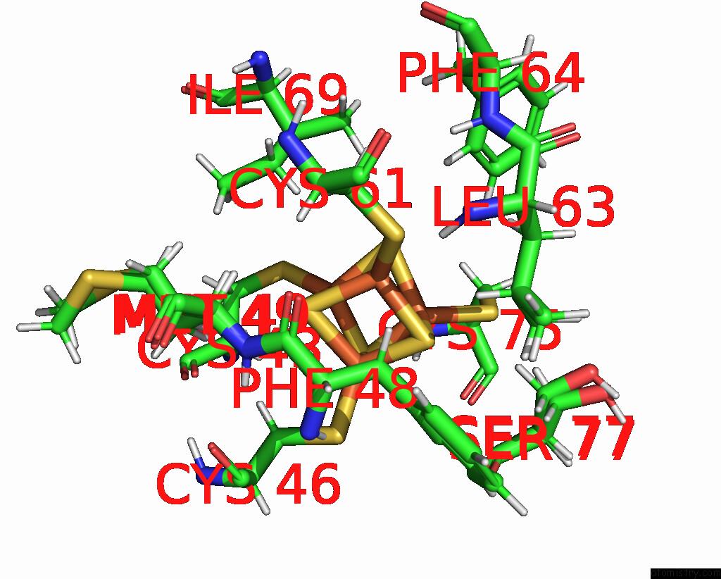
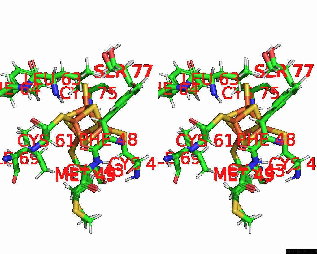
Iron binding site 1 out of 4 in 7vos
Go back to
Iron binding site 1 out
of 4 in the High-Resolution Neutron and X-Ray Joint Refined Structure of High- Potential Iron-Sulfur Protein in the Oxidized State
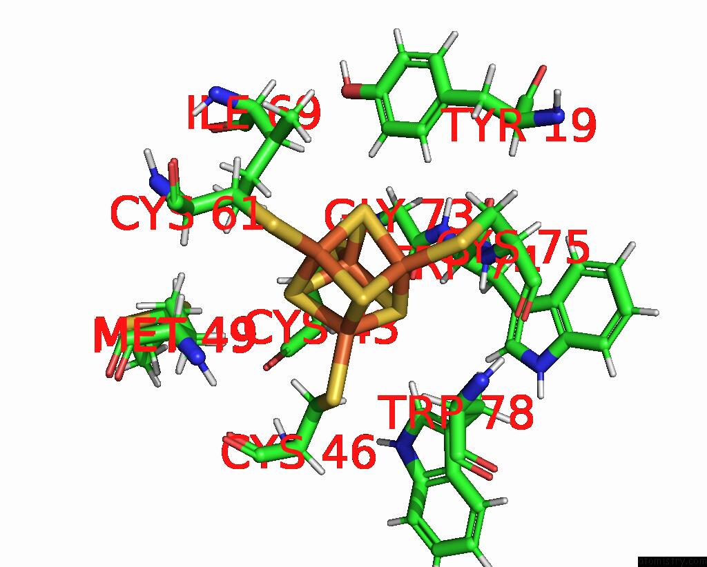
Mono view
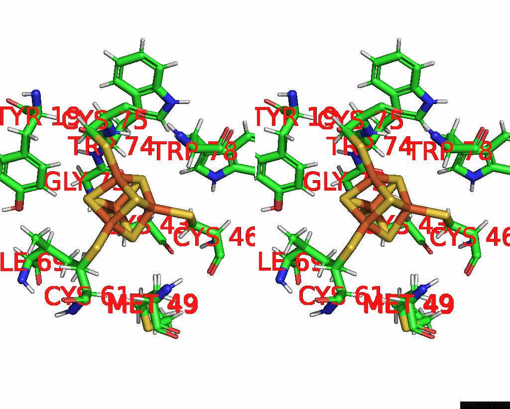
Stereo pair view
Mono view
Stereo pair view
A full contact list of Iron with other atoms in the Fe binding
site number 1 of High-Resolution Neutron and X-Ray Joint Refined Structure of High- Potential Iron-Sulfur Protein in the Oxidized State within 5.0Å range:
|
Iron binding site 2 out of 4 in 7vos
Go back to
Iron binding site 2 out
of 4 in the High-Resolution Neutron and X-Ray Joint Refined Structure of High- Potential Iron-Sulfur Protein in the Oxidized State
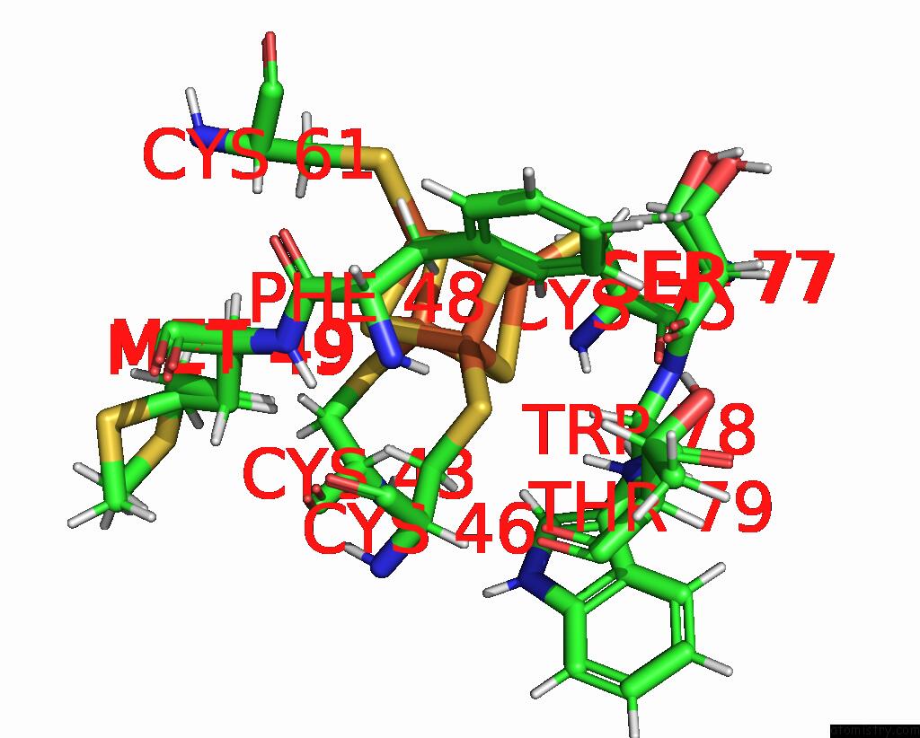
Mono view
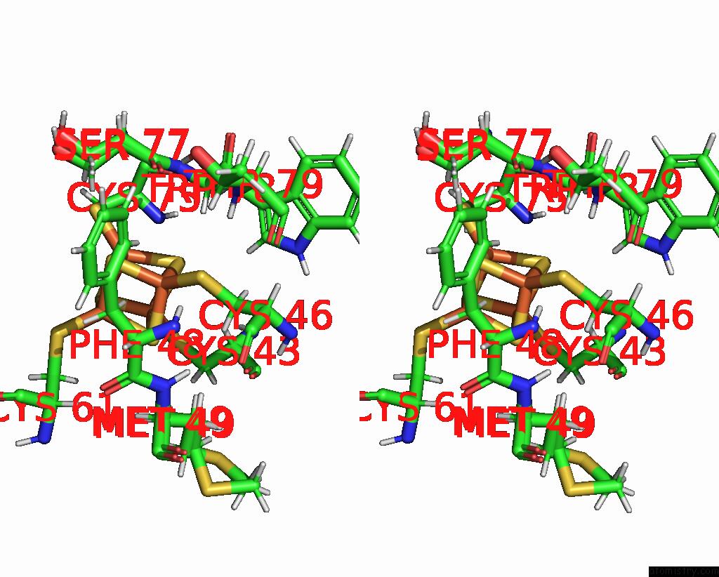
Stereo pair view
Mono view
Stereo pair view
A full contact list of Iron with other atoms in the Fe binding
site number 2 of High-Resolution Neutron and X-Ray Joint Refined Structure of High- Potential Iron-Sulfur Protein in the Oxidized State within 5.0Å range:
|
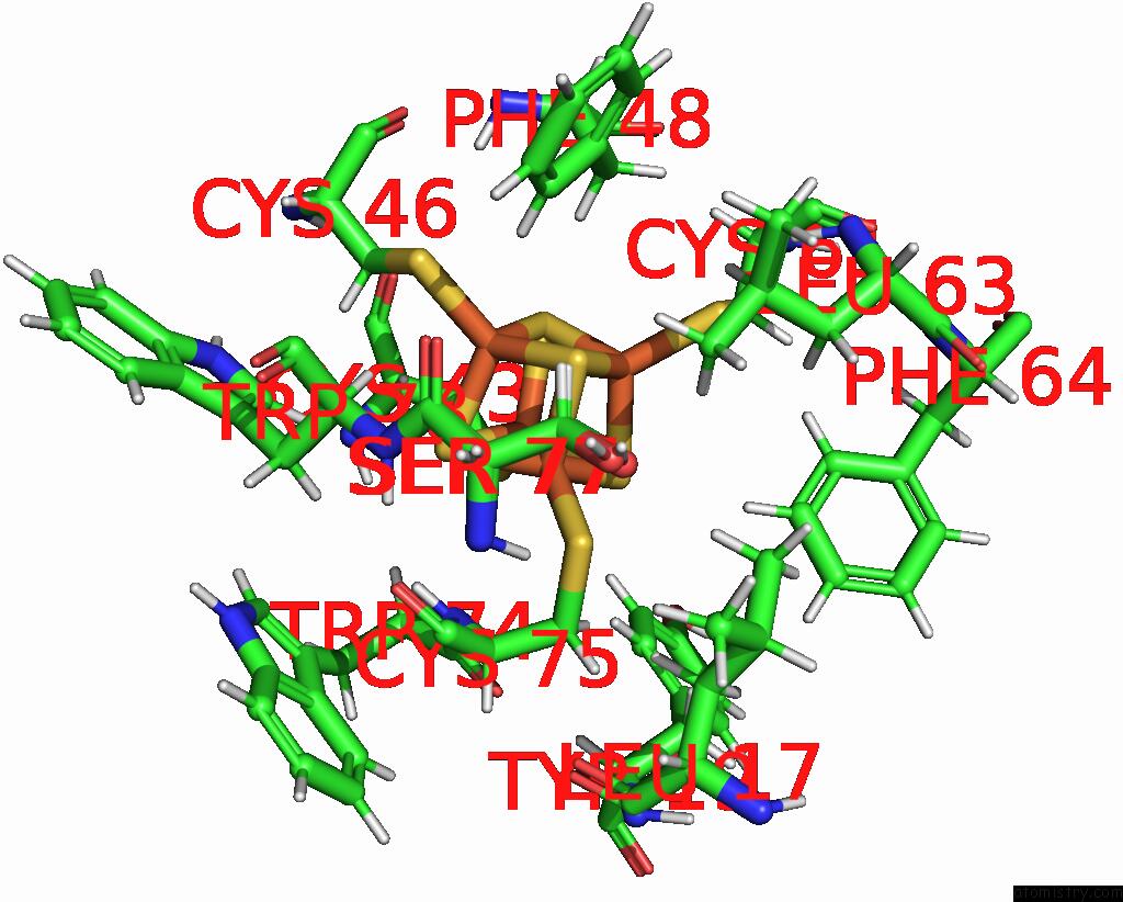
Iron binding site 3 out of 4 in 7vos
Go back to
Iron binding site 3 out
of 4 in the High-Resolution Neutron and X-Ray Joint Refined Structure of High- Potential Iron-Sulfur Protein in the Oxidized State
Mono view
Stereo pair view
Mono view
Stereo pair view
A full contact list of Iron with other atoms in the Fe binding
site number 3 of High-Resolution Neutron and X-Ray Joint Refined Structure of High- Potential Iron-Sulfur Protein in the Oxidized State within 5.0Å range:
|
Iron binding site 4 out of 4 in 7vos
Go back to
Iron binding site 4 out
of 4 in the High-Resolution Neutron and X-Ray Joint Refined Structure of High- Potential Iron-Sulfur Protein in the Oxidized State
Mono view

Stereo pair view
Mono view
Stereo pair view
A full contact list of Iron with other atoms in the Fe binding
site number 4 of High-Resolution Neutron and X-Ray Joint Refined Structure of High- Potential Iron-Sulfur Protein in the Oxidized State within 5.0Å range:
|
Reference:
Y.Hanazono,
Y.Hirano,
K.Takeda,
K.Kusaka,
T.Tamada,
K.Miki.
Revisiting the Concept of Peptide Bond Planarity in An Iron-Sulfur Protein By Neutron Structure Analysis. Sci Adv V. 8 N2276 2022.
ISSN: ESSN 2375-2548
PubMed: 35594350
DOI: 10.1126/SCIADV.ABN2276
Page generated: Fri Aug 9 05:44:00 2024
ISSN: ESSN 2375-2548
PubMed: 35594350
DOI: 10.1126/SCIADV.ABN2276
Last articles
Cl in 3A7KCl in 3A6R
Cl in 3A7S
Cl in 3A6Q
Cl in 3A6N
Cl in 3A6L
Cl in 3A6K
Cl in 3A6H
Cl in 3A4P
Cl in 363D