Iron »
PDB 7vp8-7w1p »
7vp8 »
Iron in PDB 7vp8: Crystal Structure of Ferritin From Ureaplasma Urealyticum
Protein crystallography data
The structure of Crystal Structure of Ferritin From Ureaplasma Urealyticum, PDB code: 7vp8
was solved by
W.Wang,
X.Liu,
Y.Wang,
D.Fu,
H.Wang,
with X-Ray Crystallography technique. A brief refinement statistics is given in the table below:
| Resolution Low / High (Å) | 34.34 / 2.00 |
| Space group | F 4 3 2 |
| Cell size a, b, c (Å), α, β, γ (°) | 178.441, 178.441, 178.441, 90, 90, 90 |
| R / Rfree (%) | 15.9 / 19.1 |
Other elements in 7vp8:
The structure of Crystal Structure of Ferritin From Ureaplasma Urealyticum also contains other interesting chemical elements:
| Chlorine | (Cl) | 3 atoms |
Iron Binding Sites:
The binding sites of Iron atom in the Crystal Structure of Ferritin From Ureaplasma Urealyticum
(pdb code 7vp8). This binding sites where shown within
5.0 Angstroms radius around Iron atom.
In total 4 binding sites of Iron where determined in the Crystal Structure of Ferritin From Ureaplasma Urealyticum, PDB code: 7vp8:
Jump to Iron binding site number: 1; 2; 3; 4;
In total 4 binding sites of Iron where determined in the Crystal Structure of Ferritin From Ureaplasma Urealyticum, PDB code: 7vp8:
Jump to Iron binding site number: 1; 2; 3; 4;
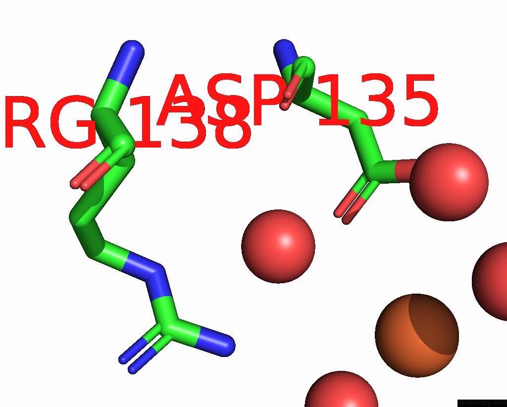
Iron binding site 1 out of 4 in 7vp8
Go back to
Iron binding site 1 out
of 4 in the Crystal Structure of Ferritin From Ureaplasma Urealyticum
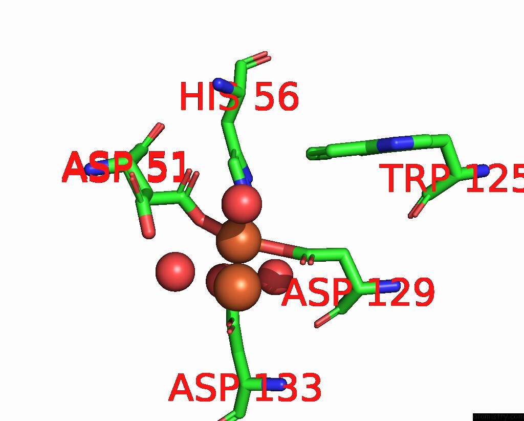
Mono view
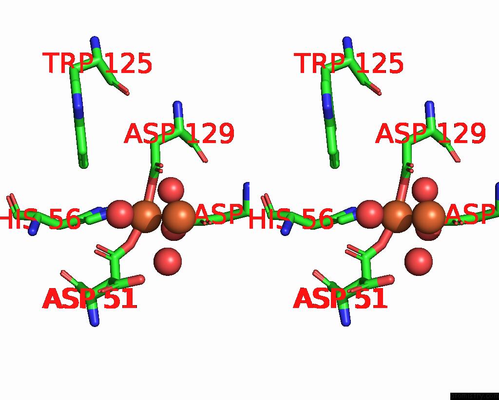
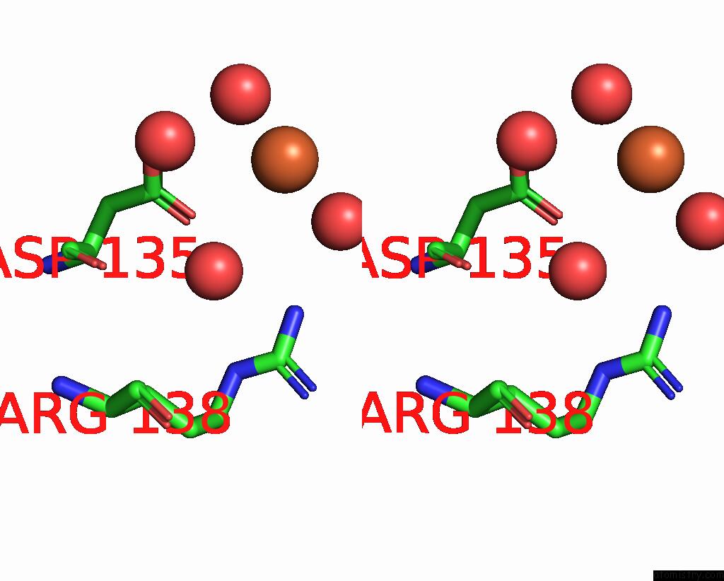
Stereo pair view
Mono view
Stereo pair view
A full contact list of Iron with other atoms in the Fe binding
site number 1 of Crystal Structure of Ferritin From Ureaplasma Urealyticum within 5.0Å range:
|
Iron binding site 2 out of 4 in 7vp8
Go back to
Iron binding site 2 out
of 4 in the Crystal Structure of Ferritin From Ureaplasma Urealyticum
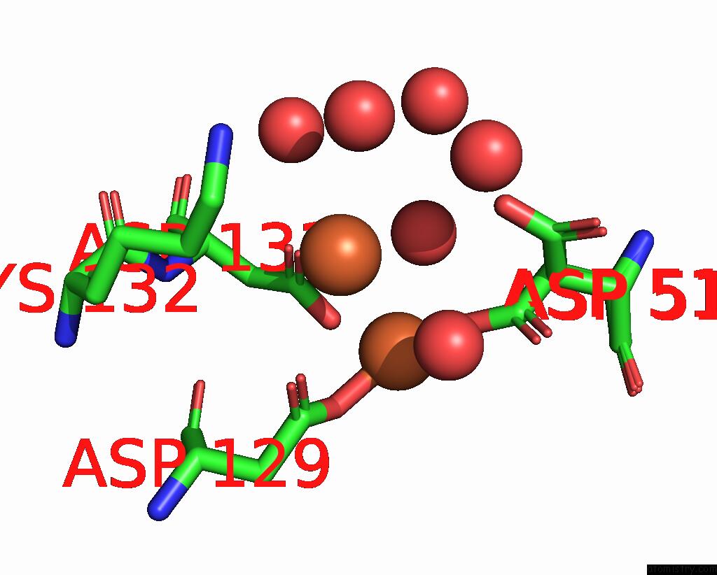
Mono view
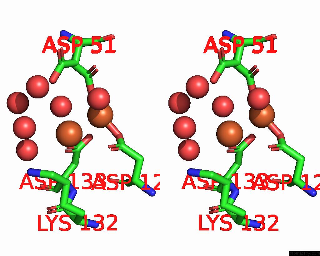
Stereo pair view
Mono view
Stereo pair view
A full contact list of Iron with other atoms in the Fe binding
site number 2 of Crystal Structure of Ferritin From Ureaplasma Urealyticum within 5.0Å range:
|
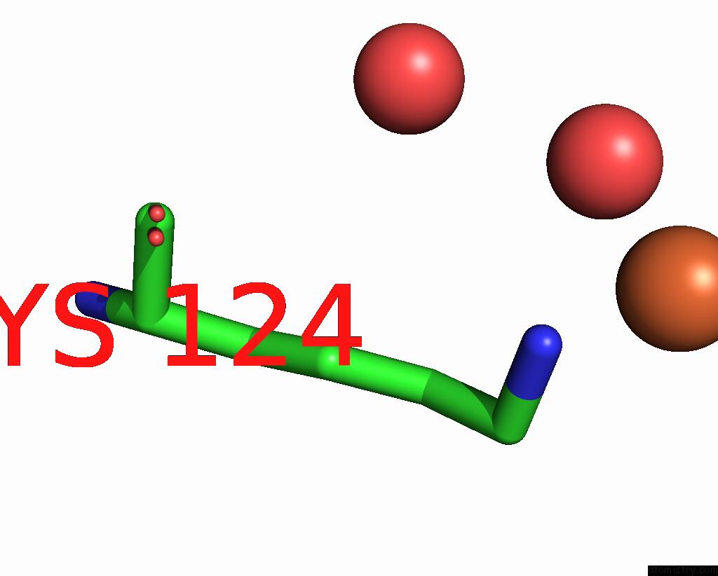
Iron binding site 3 out of 4 in 7vp8
Go back to
Iron binding site 3 out
of 4 in the Crystal Structure of Ferritin From Ureaplasma Urealyticum
Mono view
Stereo pair view
Mono view
Stereo pair view
A full contact list of Iron with other atoms in the Fe binding
site number 3 of Crystal Structure of Ferritin From Ureaplasma Urealyticum within 5.0Å range:
|
Iron binding site 4 out of 4 in 7vp8
Go back to
Iron binding site 4 out
of 4 in the Crystal Structure of Ferritin From Ureaplasma Urealyticum
Mono view

Stereo pair view
Mono view
Stereo pair view
A full contact list of Iron with other atoms in the Fe binding
site number 4 of Crystal Structure of Ferritin From Ureaplasma Urealyticum within 5.0Å range:
|
Reference:
W.Wang,
X.Liu,
Y.Wang,
Y.Wang,
D.Fu,
H.Xi,
Y.Zhao,
H.Wang.
Distinct Structural Characteristics Define A New Subfamily of Mycoplasma Ferritin Chin.Chem.Lett. V. 33 4952 2022.
ISSN: ISSN 1001-8417
DOI: 10.1016/J.CCLET.2022.03.119
Page generated: Fri Aug 9 06:02:39 2024
ISSN: ISSN 1001-8417
DOI: 10.1016/J.CCLET.2022.03.119
Last articles
F in 7QISF in 7QK4
F in 7QK0
F in 7QIR
F in 7QI8
F in 7QC5
F in 7QG3
F in 7QHN
F in 7Q6P
F in 7QBZ