Iron »
PDB 7vp8-7w1p »
7vuc »
Iron in PDB 7vuc: Myoglobin Mutant L29I/H64G/V68A
Protein crystallography data
The structure of Myoglobin Mutant L29I/H64G/V68A, PDB code: 7vuc
was solved by
K.Takahashi,
I.V.Korendovych,
J.R.H.Tame,
with X-Ray Crystallography technique. A brief refinement statistics is given in the table below:
| Resolution Low / High (Å) | 22.68 / 1.40 |
| Space group | P 6 |
| Cell size a, b, c (Å), α, β, γ (°) | 90.019, 90.019, 45.368, 90, 90, 120 |
| R / Rfree (%) | 17.6 / 19.5 |
Iron Binding Sites:
The binding sites of Iron atom in the Myoglobin Mutant L29I/H64G/V68A
(pdb code 7vuc). This binding sites where shown within
5.0 Angstroms radius around Iron atom.
In total only one binding site of Iron was determined in the Myoglobin Mutant L29I/H64G/V68A, PDB code: 7vuc:
In total only one binding site of Iron was determined in the Myoglobin Mutant L29I/H64G/V68A, PDB code: 7vuc:
Iron binding site 1 out of 1 in 7vuc
Go back to
Iron binding site 1 out
of 1 in the Myoglobin Mutant L29I/H64G/V68A
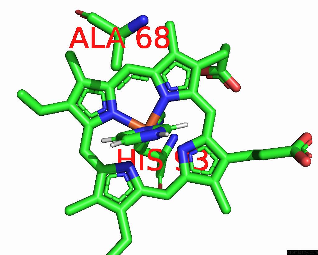
Mono view
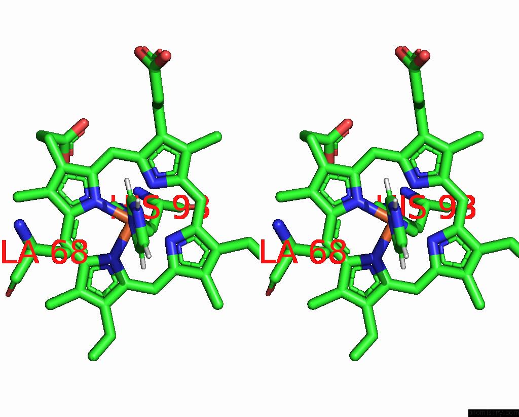
Stereo pair view
Mono view
Stereo pair view
A full contact list of Iron with other atoms in the Fe binding
site number 1 of Myoglobin Mutant L29I/H64G/V68A within 5.0Å range:
|
Reference:
S.Bhattacharya,
E.G.Margheritis,
K.Takahashi,
A.Kulesha,
A.D'souza,
I.Kim,
J.H.Yoon,
J.R.H.Tame,
A.N.Volkov,
O.V.Makhlynets,
I.V.Korendovych.
uc(Nmr)-Guided Directed Evolution. Nature V. 610 389 2022.
ISSN: ESSN 1476-4687
PubMed: 36198791
DOI: 10.1038/S41586-022-05278-9
Page generated: Fri Aug 9 06:02:39 2024
ISSN: ESSN 1476-4687
PubMed: 36198791
DOI: 10.1038/S41586-022-05278-9
Last articles
Zn in 9MJ5Zn in 9HNW
Zn in 9G0L
Zn in 9FNE
Zn in 9DZN
Zn in 9E0I
Zn in 9D32
Zn in 9DAK
Zn in 8ZXC
Zn in 8ZUF