Iron »
PDB 7vqg-7w1t »
7vw4 »
Iron in PDB 7vw4: The X-Ray Structure of Sperm Whale F46C/L61C Myoglobin Double Mutant
Protein crystallography data
The structure of The X-Ray Structure of Sperm Whale F46C/L61C Myoglobin Double Mutant, PDB code: 7vw4
was solved by
Y.W.Lin,
with X-Ray Crystallography technique. A brief refinement statistics is given in the table below:
| Resolution Low / High (Å) | 24.01 / 1.80 |
| Space group | P 21 21 21 |
| Cell size a, b, c (Å), α, β, γ (°) | 39.944, 48.016, 76.847, 90, 90, 90 |
| R / Rfree (%) | 17.9 / 22.6 |
Iron Binding Sites:
The binding sites of Iron atom in the The X-Ray Structure of Sperm Whale F46C/L61C Myoglobin Double Mutant
(pdb code 7vw4). This binding sites where shown within
5.0 Angstroms radius around Iron atom.
In total only one binding site of Iron was determined in the The X-Ray Structure of Sperm Whale F46C/L61C Myoglobin Double Mutant, PDB code: 7vw4:
In total only one binding site of Iron was determined in the The X-Ray Structure of Sperm Whale F46C/L61C Myoglobin Double Mutant, PDB code: 7vw4:
Iron binding site 1 out of 1 in 7vw4
Go back to
Iron binding site 1 out
of 1 in the The X-Ray Structure of Sperm Whale F46C/L61C Myoglobin Double Mutant
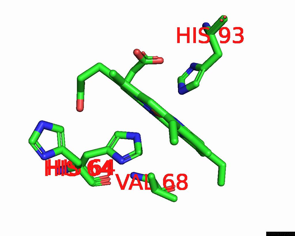
Mono view
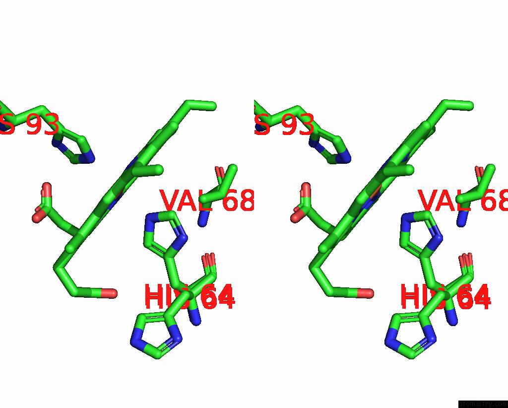
Stereo pair view
Mono view
Stereo pair view
A full contact list of Iron with other atoms in the Fe binding
site number 1 of The X-Ray Structure of Sperm Whale F46C/L61C Myoglobin Double Mutant within 5.0Å range:
|
Reference:
Y.W.Lin,
Y.W.Lin.
N/A N/A.
Page generated: Thu Aug 7 07:48:50 2025
Last articles
Mg in 1X54Mg in 1X3S
Mg in 1X1T
Mg in 1X1S
Mg in 1X1R
Mg in 1WYW
Mg in 1WVM
Mg in 1X09
Mg in 1X07
Mg in 1X06