Iron »
PDB 7vqg-7w1t »
7vyo »
Iron in PDB 7vyo: The Structure of Gdmn
Protein crystallography data
The structure of The Structure of Gdmn, PDB code: 7vyo
was solved by
J.Wei,
J.Zheng,
J.Zhou,
Q.Kang,
L.Bai,
with X-Ray Crystallography technique. A brief refinement statistics is given in the table below:
| Resolution Low / High (Å) | 36.97 / 2.25 |
| Space group | P 31 2 1 |
| Cell size a, b, c (Å), α, β, γ (°) | 110.955, 110.955, 231.544, 90, 90, 120 |
| R / Rfree (%) | 17.6 / 20.9 |
Other elements in 7vyo:
The structure of The Structure of Gdmn also contains other interesting chemical elements:
| Chlorine | (Cl) | 1 atom |
Iron Binding Sites:
The binding sites of Iron atom in the The Structure of Gdmn
(pdb code 7vyo). This binding sites where shown within
5.0 Angstroms radius around Iron atom.
In total 2 binding sites of Iron where determined in the The Structure of Gdmn, PDB code: 7vyo:
Jump to Iron binding site number: 1; 2;
In total 2 binding sites of Iron where determined in the The Structure of Gdmn, PDB code: 7vyo:
Jump to Iron binding site number: 1; 2;
Iron binding site 1 out of 2 in 7vyo
Go back to
Iron binding site 1 out
of 2 in the The Structure of Gdmn

Mono view
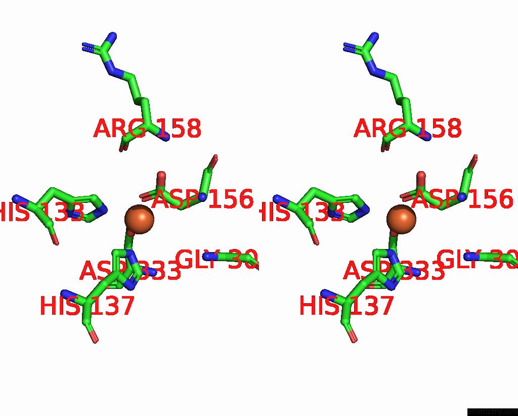
Stereo pair view
Mono view
Stereo pair view
A full contact list of Iron with other atoms in the Fe binding
site number 1 of The Structure of Gdmn within 5.0Å range:
|
Iron binding site 2 out of 2 in 7vyo
Go back to
Iron binding site 2 out
of 2 in the The Structure of Gdmn
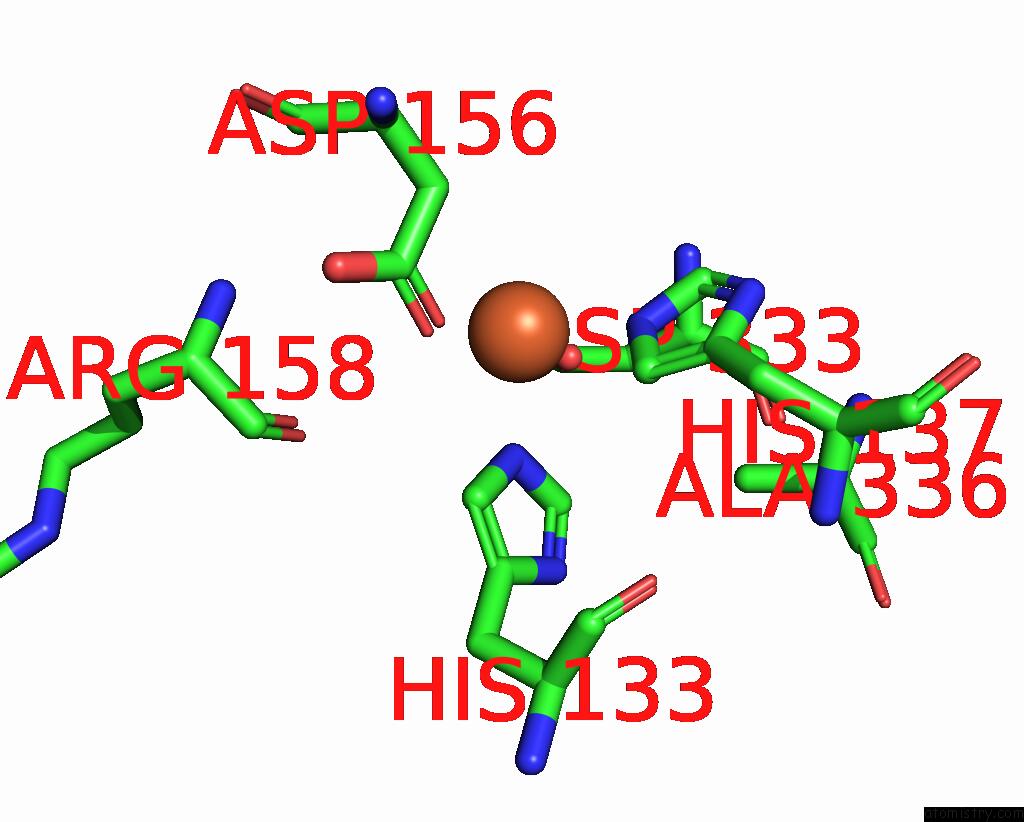
Mono view
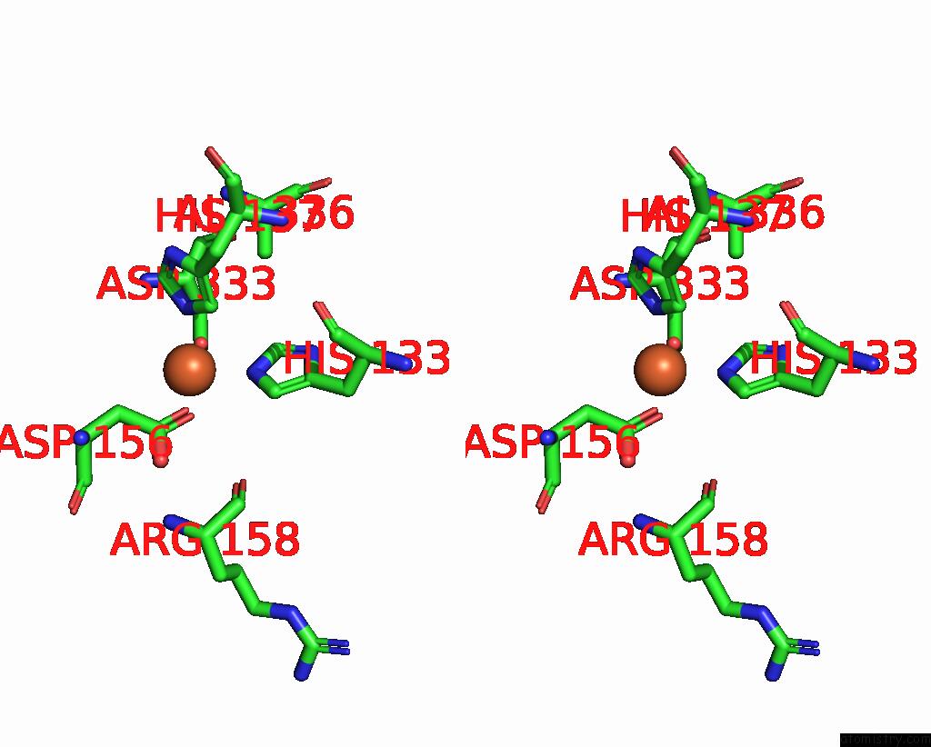
Stereo pair view
Mono view
Stereo pair view
A full contact list of Iron with other atoms in the Fe binding
site number 2 of The Structure of Gdmn within 5.0Å range:
|
Reference:
J.Wei,
X.Zhang,
Y.Zhou,
X.Cheng,
Z.Lin,
M.Tang,
J.Zheng,
B.Wang,
Q.Kang,
L.Bai.
Endowing Homodimeric Carbamoyltransferase Gdmn with Iterative Functions Through Structural Characterization and Mechanistic Studies. Nat Commun V. 13 6617 2022.
ISSN: ESSN 2041-1723
PubMed: 36329057
DOI: 10.1038/S41467-022-34387-2
Page generated: Thu Aug 7 08:14:46 2025
ISSN: ESSN 2041-1723
PubMed: 36329057
DOI: 10.1038/S41467-022-34387-2
Last articles
Mg in 2J59Mg in 2J4L
Mg in 2J4E
Mg in 2J4Q
Mg in 2J4K
Mg in 2J4H
Mg in 2J4J
Mg in 2J3D
Mg in 2J3Q
Mg in 2J3E