Iron »
PDB 7w1u-7wdg »
7w8f »
Iron in PDB 7w8f: Crystal Structure of Siderophore Binding Protein Vatd From Vibrio Vulnificus M2799 Complexed with Desferal
Protein crystallography data
The structure of Crystal Structure of Siderophore Binding Protein Vatd From Vibrio Vulnificus M2799 Complexed with Desferal, PDB code: 7w8f
was solved by
K.Tomoo,
K.Miyamoto,
with X-Ray Crystallography technique. A brief refinement statistics is given in the table below:
| Resolution Low / High (Å) | 26.10 / 1.90 |
| Space group | P 1 21 1 |
| Cell size a, b, c (Å), α, β, γ (°) | 34.8, 57.89, 62.7, 90, 95.02, 90 |
| R / Rfree (%) | 17.7 / 21.9 |
Other elements in 7w8f:
The structure of Crystal Structure of Siderophore Binding Protein Vatd From Vibrio Vulnificus M2799 Complexed with Desferal also contains other interesting chemical elements:
| Magnesium | (Mg) | 2 atoms |
Iron Binding Sites:
The binding sites of Iron atom in the Crystal Structure of Siderophore Binding Protein Vatd From Vibrio Vulnificus M2799 Complexed with Desferal
(pdb code 7w8f). This binding sites where shown within
5.0 Angstroms radius around Iron atom.
In total 2 binding sites of Iron where determined in the Crystal Structure of Siderophore Binding Protein Vatd From Vibrio Vulnificus M2799 Complexed with Desferal, PDB code: 7w8f:
Jump to Iron binding site number: 1; 2;
In total 2 binding sites of Iron where determined in the Crystal Structure of Siderophore Binding Protein Vatd From Vibrio Vulnificus M2799 Complexed with Desferal, PDB code: 7w8f:
Jump to Iron binding site number: 1; 2;
Iron binding site 1 out of 2 in 7w8f
Go back to
Iron binding site 1 out
of 2 in the Crystal Structure of Siderophore Binding Protein Vatd From Vibrio Vulnificus M2799 Complexed with Desferal
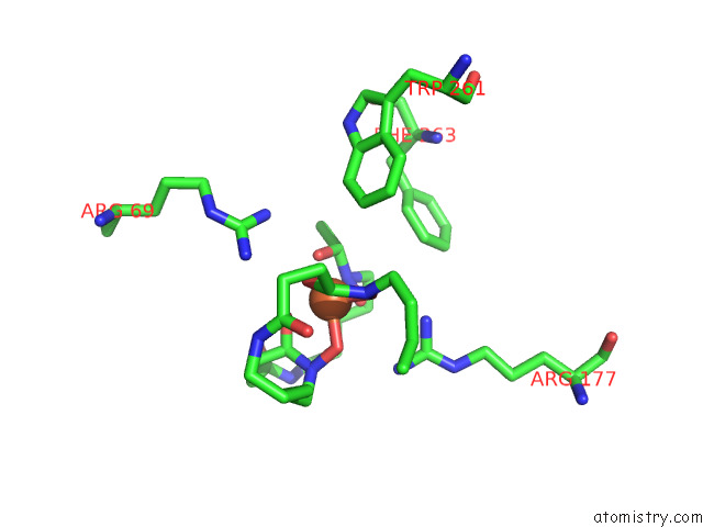
Mono view
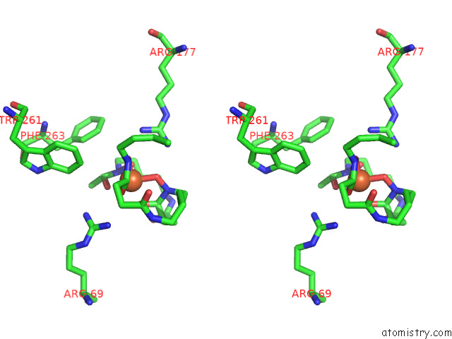
Stereo pair view
Mono view
Stereo pair view
A full contact list of Iron with other atoms in the Fe binding
site number 1 of Crystal Structure of Siderophore Binding Protein Vatd From Vibrio Vulnificus M2799 Complexed with Desferal within 5.0Å range:
|
Iron binding site 2 out of 2 in 7w8f
Go back to
Iron binding site 2 out
of 2 in the Crystal Structure of Siderophore Binding Protein Vatd From Vibrio Vulnificus M2799 Complexed with Desferal
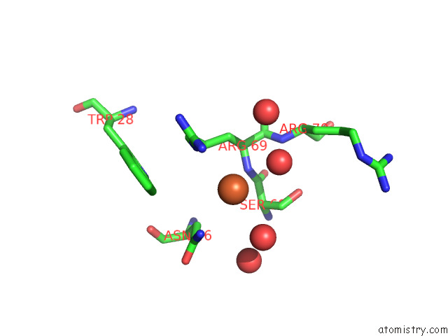
Mono view
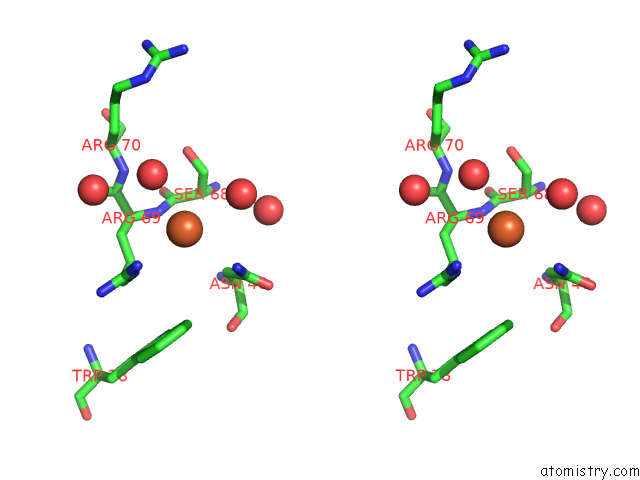
Stereo pair view
Mono view
Stereo pair view
A full contact list of Iron with other atoms in the Fe binding
site number 2 of Crystal Structure of Siderophore Binding Protein Vatd From Vibrio Vulnificus M2799 Complexed with Desferal within 5.0Å range:
|
Reference:
K.Tomoo,
K.Miyamoto.
Crystal Structure of Siderophore Binding Protein Vatd From Vibrio Vulnificus M2799 Complexed with Desferal To Be Published.
Page generated: Thu Aug 7 09:47:54 2025
Last articles
Mg in 1VQ4Mg in 1VPA
Mg in 1VPE
Mg in 1VOM
Mg in 1VMA
Mg in 1VMK
Mg in 1VM9
Mg in 1VCR
Mg in 1VLB
Mg in 1VKP